Central Auckland, East Auckland, North Auckland, South Auckland, West Auckland > Private Hospitals & Specialists >
Oral Surgery Associates
Private Service, Oral & Maxillofacial Surgery
Today
Description
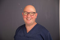
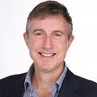
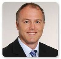
Oral Surgery Associates, originally established by Neil Luyk (now retired) is the exciting venture of three Oral & Maxillofacial Surgeons, expert in surgery of the mouth and face.
A comprehensive oral and maxillofacial surgery (surgery of the mouth, face and jaws) service is available, and all aspects of oral and maxillofacial surgery are provided. The service includes comprehensive management of:
- dento-alveolar problems such as impacted wisdom teeth,
- oral medicine problems such as oral ulceration,
- facial reconstructive surgery including corrective jaw surgery (orthognathic surgery),
- dental reconstructive surgery with dental implants.
The surgeons are all Southern Cross Affliated Providers.
We have recently added to our surgical armamentarium with the purchase of two new medical lasers and can now offer oral hard and soft tissue laser surgery with significantly less post-operative pain and swelling following surgery.
Consultants
-

Dr Ian Cathro
Oral & Maxillofacial Surgeon
-

Dr John Harrison
Oral & Maxillofacial Surgeon
-

Dr Cameron Lewis
Oral & Maxillofacial Surgeon
How do I access this service?
Referral Expectations
Most patients are referred by their doctor or dentist but it is not a necessity.
Fees and Charges Categorisation
Fees apply
Fees and Charges Description
The surgeons at Oral Surgery Associates are Southern Cross Affiliated Provider for procedures under the Oral/maxillofacial surgery (Mouth/teeth/gum/jaw surgery) service category. These include :
- Apicectomy
- Arthrocentesis
- Cone beam CT
- Consultation
- Extraction of wisdom teeth
- Frenectomy
- Oral biopsies
- Pericision
Hours
| Mon – Thu | 8:00 AM – 4:00 PM |
|---|---|
| Fri | 9:00 AM – 3:30 PM |
Public Holidays: Closed Good Friday (18 Apr), Easter Sunday (20 Apr), Easter Monday (21 Apr), ANZAC Day (25 Apr), King's Birthday (2 Jun), Matariki (20 Jun), Labour Day (27 Oct), Auckland Anniversary (26 Jan), Waitangi Day (6 Feb).
Services Provided
Gum tissue at the site of the implant is opened up to expose the bone. The bone is drilled and a titanium implant is inserted where the root of your tooth had been. Once the bone and gum has healed (3-6 months), the post is attached to the implant and the crown is placed over the post and cemented into place.
Gum tissue at the site of the implant is opened up to expose the bone. The bone is drilled and a titanium implant is inserted where the root of your tooth had been. Once the bone and gum has healed (3-6 months), the post is attached to the implant and the crown is placed over the post and cemented into place.
Gum tissue at the site of the implant is opened up to expose the bone. The bone is drilled and a titanium implant is inserted where the root of your tooth had been. Once the bone and gum has healed (3-6 months), the post is attached to the implant and the crown is placed over the post and cemented into place.
Parotidectomy: an incision (cut) is made in front of the ear and runs down below the jaw line. Part or all of the parotid gland is removed. Superficial parotidectomy: an incision is made in front of the ear and runs down beneath the ear lobe. The superficial (top) lobe of the parotid gland is removed. Submandibular gland surgery: an incision is made just below the jaw bone and the submandibular gland removed.
Parotidectomy: an incision (cut) is made in front of the ear and runs down below the jaw line. Part or all of the parotid gland is removed. Superficial parotidectomy: an incision is made in front of the ear and runs down beneath the ear lobe. The superficial (top) lobe of the parotid gland is removed. Submandibular gland surgery: an incision is made just below the jaw bone and the submandibular gland removed.
Parotidectomy: an incision (cut) is made in front of the ear and runs down below the jaw line. Part or all of the parotid gland is removed.
Superficial parotidectomy: an incision is made in front of the ear and runs down beneath the ear lobe. The superficial (top) lobe of the parotid gland is removed.
Submandibular gland surgery: an incision is made just below the jaw bone and the submandibular gland removed.
Wisdom teeth are the third molars right at the back of your mouth. They usually appear during your late teens or early twenties. If there is not enough room in your mouth they may partially erupt through the gum or not at all. This is referred to as an impacted wisdom tooth. Due to their location wisdom teeth can be difficult to clean and are more susceptible to decay, gum disease and recurrent infections. They can cause crowding of teeth and, on rare occasions, cysts and tumours develop around them. Your dentist will advise if some or all of your wisdom teeth need to be removed. Wisdom teeth will usually only be removed if your dentist believes they will be a significant compromise to your oral health. Impacted tooth extraction Your dentist may recommend extraction if you are at significantly greater risk of infection or tooth decay. Impacted teeth may be removed by your dentist or they may refer you to an oral & maxillofacial surgeon. An incision (cut) is made in your gum and access to the impacted tooth cleared by pushing aside gum tissue and, if necessary, removing some bone. The tooth is removed whole or in pieces and the gum stitched together over the hole.
Wisdom teeth are the third molars right at the back of your mouth. They usually appear during your late teens or early twenties. If there is not enough room in your mouth they may partially erupt through the gum or not at all. This is referred to as an impacted wisdom tooth. Due to their location wisdom teeth can be difficult to clean and are more susceptible to decay, gum disease and recurrent infections. They can cause crowding of teeth and, on rare occasions, cysts and tumours develop around them. Your dentist will advise if some or all of your wisdom teeth need to be removed. Wisdom teeth will usually only be removed if your dentist believes they will be a significant compromise to your oral health. Impacted tooth extraction Your dentist may recommend extraction if you are at significantly greater risk of infection or tooth decay. Impacted teeth may be removed by your dentist or they may refer you to an oral & maxillofacial surgeon. An incision (cut) is made in your gum and access to the impacted tooth cleared by pushing aside gum tissue and, if necessary, removing some bone. The tooth is removed whole or in pieces and the gum stitched together over the hole.
Wisdom teeth are the third molars right at the back of your mouth. They usually appear during your late teens or early twenties. If there is not enough room in your mouth they may partially erupt through the gum or not at all. This is referred to as an impacted wisdom tooth.
Due to their location wisdom teeth can be difficult to clean and are more susceptible to decay, gum disease and recurrent infections. They can cause crowding of teeth and, on rare occasions, cysts and tumours develop around them.
Your dentist will advise if some or all of your wisdom teeth need to be removed. Wisdom teeth will usually only be removed if your dentist believes they will be a significant compromise to your oral health.
Impacted tooth extraction
Your dentist may recommend extraction if you are at significantly greater risk of infection or tooth decay. Impacted teeth may be removed by your dentist or they may refer you to an oral & maxillofacial surgeon.
An incision (cut) is made in your gum and access to the impacted tooth cleared by pushing aside gum tissue and, if necessary, removing some bone. The tooth is removed whole or in pieces and the gum stitched together over the hole.
Disability Assistance
Wheelchair access, Mobility parking space
Document Downloads
- Download a map of our location (PDF, 1.5 MB)
Refreshments
Just a short stroll away from Ascot Central is Quatro Cafe which will provide your family or friends with a nice place to have coffee and a bite to eat. Quatro welcome your friends and family to sit and enjoy their cafe while they wait for their loved ones.
Travel Directions
Located just off the motorway, near the Green Lane roundabout, Oral Surgery Associates can be found on the first floor of Ascot Central, 7 Ellerslie Racecourse Drive, Remuera. Ascot Central can be found opposite Ascot Hospital on the corner with Ellerslie Racecourse Drive. A sign for Fertility Associates is displayed prominently on the side of the building.
Public Transport
Oral Surgery Associates is located just off Green Lane East Road, making us very accessible for public transport users. The Auckland Transport Journey Planner will help you to plan your journey.
Parking
There is ample parking (Ascot Pay & Display Parking) conveniently located straight through the first round about when turning into Ellerslie Racecourse Drive. The first 30 minutes are free and rates are $4 per hour thereafter – please ensure you prepay for your parking and allow up to 45 minutes for your consultation appointment with us. If you hold a current mobility card there is disabled parking outside our building.
Accommodation
Ascot Central is located adjacent to the Ibis and Novotel Hotels, should you require accommodation.
Pharmacy
Within Ascot Hospital, a short walk from us - you will find Ascot Pharmacy who will be happy to cater to your prescription needs.
Website
Contact Details
Ascot Central, 7 Ellerslie Racecourse Drive, Remuera, Auckland
Central Auckland
-
Phone
(09) 529 5910
Email
Website
Ascot Central
First Floor
7 Ellerslie Racecourse Drive
Remuera
Auckland 1051
Street Address
Ascot Central
First Floor
7 Ellerslie Racecourse Drive
Remuera
Auckland 1051
Postal Address
Ascot Central
First Floor
7 Ellerslie Racecourse Drive
Remuera
Auckland 1051
Was this page helpful?
This page was last updated at 10:40AM on March 13, 2025. This information is reviewed and edited by Oral Surgery Associates.

