North Auckland, West Auckland > Private Hospitals & Specialists > Southern Cross Hospitals >
Southern Cross North Harbour Hospital - Orthopaedic Surgery
Private Surgical Service, Orthopaedics
Description
Established in 1991, our North Harbour Hospital typically provides services to over 7,000 patients annually, and is widely known amongst patients and specialists in the area. It is their demand for our services that drives new investments, and that has delivered significant extensions, implementation of new technologies, and ongoing improvements to operating and nursing facilities. Recent upgrades have also seen improvements to recovery and day stay and pre-admission facilities and specialist consulting areas.
Within the grounds of the North Harbour Hospital campus is the Northern Clinic, which comprises Specialist consulting, Endoscopy services, Image Guided Healthcare, Radiology services and rehabilitation.
The hospital campus offers specialists and their patients access to new advanced operating theatres, including a robotically-assisted surgical system and other advanced technologies to support advanced imaging and a range of minimally invasive treatments. New recovery areas and other patient facilities have also been added, during 2017, along with new consulting rooms and a purpose-built endoscopy suite. These facilities offer patients a wide range of specialist services on one developing health campus which also includes a pharmacy, radiology unit, cardiology services, and a specially designed post-operative intermediate care facility - which helps to increase the scope and complexity of surgical services that can be provided within the private sector on Auckland's North Shore.
Consultants
-
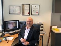
Mr Ali Bayan
Orthopaedic Surgeon
-
Mr Hugh Blackley
Orthopaedic Surgeon
-

Mr Matthew Boyle
Orthopaedic Surgeon
-
Mr Matthew Brick
Orthopaedic Surgeon
-

Mr Anthony Cheng
Orthopaedic Surgeon
-
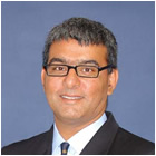
Mr Simon Chinchanwala
Orthopaedic Surgeon
-
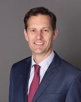
Mr Robert Elliott
Orthopaedic Surgeon
-

Mr Bill Farrington
Orthopaedic Surgeon
-
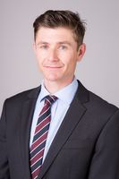
Mr Phillip Insull
Orthopaedic Surgeon
-
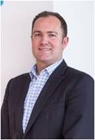
Mr Warren Leigh
Orthopaedic Surgeon
-
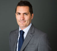
Mr Peter Misur
Orthopaedic Surgeon
-

Mr Paul Monk
Orthopaedic Surgeon
-

Mr Peter Mutch
Orthopaedic Surgeon
-
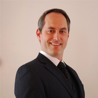
Mr John Mutu-Grigg
Orthopaedic Surgeon
-
Mr Peter Poon
Orthopaedic Surgeon
-
Mr Dean Schluter
Orthopaedic Surgeon
-
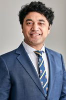
Mr Joshua Sevao
Orthopaedic Surgeon
-

Mr Rob Sharp
Orthopaedic Surgeon
-
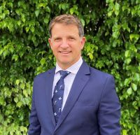
Mr Eric Swanton
Orthopaedic Surgeon
-
Mr Rupert Van Rooyen
Orthopaedic Surgeon
-

Mr Matt Walker
Orthopaedic Surgeon
-
Mr Edward Yee
Orthopaedic Surgeon
-
Mr Albert Yoon
Orthopaedic Surgeon
-
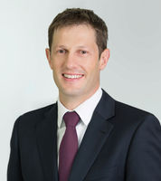
Mr Simon Young
Orthopaedic Surgeon
Procedures / Treatments
Two or three small incisions (cuts) are made in the ankle and a small telescopic instrument with a tiny camera attached (arthroscope) is inserted. This allows the surgeon to look inside the joint, identify problems and, in some cases, operate. Tiny instruments can be passed through the arthroscope to remove bony spurs, damaged cartilage or inflamed tissue.
Two or three small incisions (cuts) are made in the ankle and a small telescopic instrument with a tiny camera attached (arthroscope) is inserted. This allows the surgeon to look inside the joint, identify problems and, in some cases, operate. Tiny instruments can be passed through the arthroscope to remove bony spurs, damaged cartilage or inflamed tissue.
Two or three small incisions (cuts) are made in the ankle and a small telescopic instrument with a tiny camera attached (arthroscope) is inserted. This allows the surgeon to look inside the joint, identify problems and, in some cases, operate. Tiny instruments can be passed through the arthroscope to remove bony spurs, damaged cartilage or inflamed tissue.
An incision (cut) is made in the front of, and several smaller cuts on the outside of, the ankle. The damaged ankle joint is replaced with a metal and plastic implant.
An incision (cut) is made in the front of, and several smaller cuts on the outside of, the ankle. The damaged ankle joint is replaced with a metal and plastic implant.
An incision (cut) is made in the front of, and several smaller cuts on the outside of, the ankle. The damaged ankle joint is replaced with a metal and plastic implant.
Carpal Tunnel Syndrome is caused by a pinched nerve in the wrist that causes tingling, numbness and pain in your hand. Surgery to relieve carpal tunnel syndrome involves making an incision (cut) from the middle of the palm of your hand to your wrist. Tissue that is pressing on the nerve is then cut to release the pressure.
Carpal Tunnel Syndrome is caused by a pinched nerve in the wrist that causes tingling, numbness and pain in your hand. Surgery to relieve carpal tunnel syndrome involves making an incision (cut) from the middle of the palm of your hand to your wrist. Tissue that is pressing on the nerve is then cut to release the pressure.
Carpal Tunnel Syndrome is caused by a pinched nerve in the wrist that causes tingling, numbness and pain in your hand.
Surgery to relieve carpal tunnel syndrome involves making an incision (cut) from the middle of the palm of your hand to your wrist. Tissue that is pressing on the nerve is then cut to release the pressure.
Discectomy is an operation to remove part or all of a damaged spinal disc that is pressing on nerves, helping to relieve pain and improve movement. Microdiscectomy:a microscope is used by the surgeon to guide tiny instruments to remove the disc or disc fragments.
Discectomy is an operation to remove part or all of a damaged spinal disc that is pressing on nerves, helping to relieve pain and improve movement. Microdiscectomy:a microscope is used by the surgeon to guide tiny instruments to remove the disc or disc fragments.
Discectomy is an operation to remove part or all of a damaged spinal disc that is pressing on nerves, helping to relieve pain and improve movement.
Microdiscectomy:a microscope is used by the surgeon to guide tiny instruments to remove the disc or disc fragments.
Small incisions (cuts) are made in the hip area and a small telescopic instrument with a tiny camera attached (arthroscope) is inserted. This allows the surgeon to look inside the joint, identify problems and, in some cases, operate. Tiny instruments can be passed through the arthroscope to remove loose, damaged or inflamed tissue.
Small incisions (cuts) are made in the hip area and a small telescopic instrument with a tiny camera attached (arthroscope) is inserted. This allows the surgeon to look inside the joint, identify problems and, in some cases, operate. Tiny instruments can be passed through the arthroscope to remove loose, damaged or inflamed tissue.
Small incisions (cuts) are made in the hip area and a small telescopic instrument with a tiny camera attached (arthroscope) is inserted. This allows the surgeon to look inside the joint, identify problems and, in some cases, operate. Tiny instruments can be passed through the arthroscope to remove loose, damaged or inflamed tissue.
An incision (cut) is made on the side of the thigh to allow the surgeon access to the hip joint. The diseased and damaged parts of the hip joint are removed and replaced with smooth, artificial metal ‘ball’ and plastic ‘socket’ parts.
An incision (cut) is made on the side of the thigh to allow the surgeon access to the hip joint. The diseased and damaged parts of the hip joint are removed and replaced with smooth, artificial metal ‘ball’ and plastic ‘socket’ parts.
An incision (cut) is made on the side of the thigh to allow the surgeon access to the hip joint. The diseased and damaged parts of the hip joint are removed and replaced with smooth, artificial metal ‘ball’ and plastic ‘socket’ parts.
Several small incisions (cuts) are made on the knee through which is inserted a small telescopic instrument with a tiny camera attached (arthroscope). This allows the surgeon to look inside the joint, identify problems and, in some cases, make repairs to damaged tissue.
Several small incisions (cuts) are made on the knee through which is inserted a small telescopic instrument with a tiny camera attached (arthroscope). This allows the surgeon to look inside the joint, identify problems and, in some cases, make repairs to damaged tissue.
Several small incisions (cuts) are made on the knee through which is inserted a small telescopic instrument with a tiny camera attached (arthroscope). This allows the surgeon to look inside the joint, identify problems and, in some cases, make repairs to damaged tissue.
An incision (cut) is made on the front of the knee to allow the surgeon access to the knee joint. The damaged and painful areas of the thigh bone (femur) and lower leg bone (tibia), including the knee joint, are removed and replaced with metal and plastic parts.
An incision (cut) is made on the front of the knee to allow the surgeon access to the knee joint. The damaged and painful areas of the thigh bone (femur) and lower leg bone (tibia), including the knee joint, are removed and replaced with metal and plastic parts.
An incision (cut) is made on the front of the knee to allow the surgeon access to the knee joint. The damaged and painful areas of the thigh bone (femur) and lower leg bone (tibia), including the knee joint, are removed and replaced with metal and plastic parts.
Several small incisions (cuts) are made in the shoulder through which is inserted a small telescopic instrument with a tiny camera attached (arthroscope). The surgeon is then able to remove any bony spurs or inflamed tissue and mend torn tendons of the rotator cuff group.
Several small incisions (cuts) are made in the shoulder through which is inserted a small telescopic instrument with a tiny camera attached (arthroscope). The surgeon is then able to remove any bony spurs or inflamed tissue and mend torn tendons of the rotator cuff group.
Several small incisions (cuts) are made in the shoulder through which is inserted a small telescopic instrument with a tiny camera attached (arthroscope). The surgeon is then able to remove any bony spurs or inflamed tissue and mend torn tendons of the rotator cuff group.
This surgery involves making several small incisions (cuts) on the shoulder through which is inserted a small telescopic instrument with a tiny camera attached (arthroscope). This allows the surgeon to look inside the shoulder, identify problems and, in some cases, make repairs to damaged tissue.
This surgery involves making several small incisions (cuts) on the shoulder through which is inserted a small telescopic instrument with a tiny camera attached (arthroscope). This allows the surgeon to look inside the shoulder, identify problems and, in some cases, make repairs to damaged tissue.
This surgery involves making several small incisions (cuts) on the shoulder through which is inserted a small telescopic instrument with a tiny camera attached (arthroscope). This allows the surgeon to look inside the shoulder, identify problems and, in some cases, make repairs to damaged tissue.
An incision (cut) is made over the relevant part of the spine. Two or more vertebrae (the small bones that make up the spinal column) are fused together with bone grafts and/or metal rods to form a single bone.
An incision (cut) is made over the relevant part of the spine. Two or more vertebrae (the small bones that make up the spinal column) are fused together with bone grafts and/or metal rods to form a single bone.
An incision (cut) is made over the relevant part of the spine. Two or more vertebrae (the small bones that make up the spinal column) are fused together with bone grafts and/or metal rods to form a single bone.
An incision (cut) is made over the damaged tendon. The damaged ends of the tendon are sewn together and, if necessary, reattached to surrounding tissue.
An incision (cut) is made over the damaged tendon. The damaged ends of the tendon are sewn together and, if necessary, reattached to surrounding tissue.
An incision (cut) is made over the damaged tendon. The damaged ends of the tendon are sewn together and, if necessary, reattached to surrounding tissue.
Visiting Hours
- Weekdays: 11:00 to 20:00
- Weekends: 11:00 to 20:00
Contact Details
Southern Cross North Harbour Hospital,
North Auckland
Phone: (09) 925 4400
Fax: (09) 925 4434
E-mail: northharbour@southerncrosshospitals.co.nz
Location details here
Surgeons can be contacted directly at their private consultation rooms.
232 Wairau Road
Totara Vale
Kaipātiki
Auckland 0629
Street Address
232 Wairau Road
Totara Vale
Kaipātiki
Auckland 0629
Postal Address
P.O. Box 101-488,
North Shore Mail Centre,
Auckland City, 0745
Was this page helpful?
This page was last updated at 2:35PM on October 3, 2024. This information is reviewed and edited by Southern Cross North Harbour Hospital - Orthopaedic Surgery.

