South Auckland > Public Hospital Services >
Auckland Regional Plastic Reconstructive and Hand Surgery
Public Service, Plastic Surgery, Orthopaedics
Description
Plastic surgery covers a wide range of different surgical procedures that repair, reconstruct or replace structures in many different parts of the body including the skin, face and head, hands, breast and stomach. Plastic surgery does not involve the use of plastic materials.
Reconstructive plastic surgery is performed on parts of the body that are abnormal or have been affected by a birth defect, accident or disease. This includes cleft lip and palate repair, scar revision or reconstruction (including skin grafts) following burns. Surgery is usually performed to improve function, but may also be performed to bring the appearance of a part of the body as close as possible to normal.
Plastic Reconstructive and Hand Surgery Services Provided by Counties Manukau Health
Counties Manukau Health Plastic Reconstructive and Hand Surgery services are located in South Auckland at Middlemore Hospital and Manukau SuperClinic™.
The Plastic Reconstructive and Hand Surgery service at CMDHB is one of the four regional plastic surgery centres in New Zealand and provides services to people who live in the Counties Manukau, Auckland, Waitemata and Northland DHB areas.
New Zealand doctors now diagnose nearly 70,000 skin cancers each year with some patients waiting many months for treatment at a public hospital. The key benefits of this new model of care mean suitable patients will be able to have their treatment at their first appointment. Other patients with some common hand problems such as carpal tunnel syndrome will also be able to have their surgery performed under local anaesthetic on the same basis.
This See and Treat facility staff includes specialists from the Department of Plastic Surgery as well as junior doctors and primary care providers such as local general practitioners (GPs). This collaborative model utilising GPs within the public healthcare system will promote higher skill development within primary care providers, with the goal to one day open multiple effective locality hubs where trained GPs can treat patients closer to home. Building stronger relationships between GPs and DHB consultant surgeons will be an additional benefit of this initiative.
All patient referrals sent to the Auckland Regional Centre for Plastic Reconstructive & Hand Surgery will be screened for suitability. Plastic Surgery clinics will continue to run at Manukau SuperClinic for other medical conditions, but if the patient is deemed appropriate, they will be called into the See and Treat Unit at Middlemore Hospital instead. This will significantly improve the service provided to our patients.
This facility to see and treat is located at Middlemore Hospital, Level 1 of the Galbraith Building.
If you have any questions please feel free to email the Service Manager Crisna Taljaard (CMDHB)
Consultants
-

Mr Paul Baker
Consultant Plastic Surgeon, Burn Service Clinical Leader
-
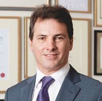
Mr Glenn Bartlett
Consultant Plastic Surgeon
-

Mr Murray Beagley
Consultant Plastic Surgeon
-

Dr Amelia Boucher
Consultant Plastic Surgeon
-
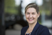
Ms Alessandra Canal
Consultant Plastic Surgeon
-

Dr Joseph Chen
Consultant Plastic Surgeon
-
Mr Ashwin Chunilal
Consultant Plastic Surgeon
-

Dr Lindsay Damkat Thomas
Consultant Plastic Surgeon
-
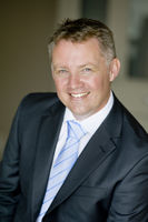
Mr Adam Durrant
Consultant Orthopaedic Hand Surgeon
-

Mr Michael Foster
Consultant Orthopaedic Hand Surgeon, Hand Service Clinical Leader
-
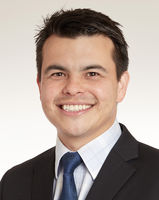
Mr Jonathan Heather
Consultant Plastic Surgeon
-

Mr Wolfgang Heiss-Dunlop
Consultant Orthopaedic Hand Surgeon
-

Ms Sarah Hulme
Consultant Plastic Surgeon
-

Mr John Kenealy
Clinical Director - SAPS, Consultant Plastic Surgeon
-

Dr Heather Le Cocq
Head of Department, Consultant Plastic Surgeon
-

Dr Victoria Lo
Consultant Plastic Surgeon
-
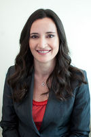
Associate Professor Michelle Locke
Deputy Head of Department, Associate Professor, Consultant Plastic Surgeon
-

Mr Thomas Maxwell
Consultant Orthopaedic Hand Surgeon
-

Mr Amber Moazzam
Consultant Plastic Surgeon
-

Mr David Morgan Jones
Consultant Plastic Surgeon
-
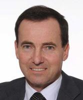
Mr Bruce Peat
Consultant Plastic Surgeon
-

Dr Andrew Sanders
Consultant Plastic Surgeon
-
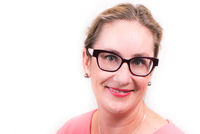
Ms Meredith Simcock
Consultant Plastic Surgeon
-

Ms Karen Smith
Consultant Orthopaedic Hand Surgeon
-
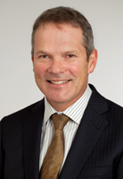
Mr Chris Taylor
Consultant Orthopaedic Hand Surgeon
-

Dr Wei Lun Wong
Consultant Plastic Surgeon
-

Mr Richard Wong She
Consultant Plastic Surgeon
Referral Expectations
Acute Patients – Middlemore Hospital
If you have an urgent problem requiring immediate surgical assessment you are referred acutely to the Emergency Department where you are initially seen by the plastic surgical junior medical staff who will decide whether you need to be admitted to Middlemore Hospital. Investigations will be performed as required, and the more senior members of the team involved where necessary.
Outpatients – Manukau SuperClinic™
If the problem is not urgent, your GP will write a letter to the Department requesting an appointment in the outpatient clinic. Waiting times for the clinics depend on the urgency of each case and vary from 2 weeks to 6 months.
You will have an initial consultation with your plastic surgeon who will assess your general health and discuss the best type of surgical procedure for you. You will be given instructions on medications to avoid before surgery.
Clinics are held in Module 5, Manukau SuperClinic™; Monday to Friday 8am - 4.30pm.
See and Treat Service – Middlemore Hospital
Covering common skin cancer and hand problems that fit within certain criteria. If suitable, your GP will write a letter to the Department requesting an appointment in our fully integrated one-stop "See and Treat" clinic, where patients could have their treatment on the same day as their first specialist appointment. Surgery will be performed under local anaesthetic and you will be given instructions on medication prior to your arrival.
Patients will be asked to make contact with their GP prior to arriving so that their GP can advise them on the management of their anticoagulants prior to their appointment.
As patients may require a biopsy or surgical procedure in the See and Treat Facility, please let us know if the patient is taking any medication to thin their blood such as warfarin, aspirin, clopidogrel, dabigatran or dipyridamole.
Clinics are held in Middlemore Hospital, Level 1 of the Galbraith Building. For further information please see your local GP or contact us for more information.
Referral is through normal referral pathway, please see information under the Referral Process
Fees and Charges Description
There are no charges for services to public patients if you are lawfully in New Zealand and meet one of the Eligibility Directions specified criteria set by the Ministry of Health. If you do not meet the criteria, you will be required to pay for the full costs of any medical treatment you receive during your stay.
To check whether you meet the specified eligibility criteria, visit the Ministry of Health website. https://www.health.govt.nz/new-zealand-health-system/eligibility-publicly-funded-health-services
CM Health has an eligibility team who can assist with any patient enquiries. If you have any queries, please phone: (09) 276 0060.
For more information, visit the Counties Manukau Health website.
Common Conditions / Procedures / Treatments
The goal of breast reconstruction surgery is to reconstruct an alternative breast mound, which resembles the form and characteristics of a normal breast. There are a number of options available and not all are suitable for every situation. Your Plastic Surgeon and team will discuss what types of reconstruction are suitable for you and when. Excess weight and smoking affect the chance of a successful outcome and therefore breast reconstruction will usually only be offered to non-smoking women with a BMI <35. Timing of Reconstruction Immediate Immediate breast reconstruction means creating the new breast mound at the same operation as your mastectomy. Delayed For various reasons some patients are not suitable, or do not wish to have a reconstruction at the time of mastectomy. Breast reconstruction can be anytime, even years after a mastectomy. Types of Breast Reconstruction There are three main types of breast reconstruction: prosthetic: uses an implant autologous: uses own tissue using a combination of both Prosthetic reconstruction: replaces the breast tissue that has been removed with an artificial implant underneath the skin and muscle of the chest wall. Autologous (own tissue) or flap reconstruction: this involves moving tissue usually skin, fat and sometimes muscle from one part of the body to create a breast shape. Combination of both: this means combining your own tissue with an implant to make a breast. There are many possible variations and different options that will be further discussed in more detail by your Plastic Surgeon. Nipple Reconstruction Nipple reconstruction is one of the final options for breast reconstruction. The nipple is created by using local breast skin or a small implant. Once this is healed it can then be tattooed to give the illusion of an areola and nipple. Most breast reconstruction operations will require several operations and clinic visits. The actual number of operations and recovery time will vary depending on what type of reconstruction you choose. These factors will be discussed with you during your consultation with your Plastic Surgeon. Breast Reconstruction Patient Information (PDF, 69.3 KB)
The goal of breast reconstruction surgery is to reconstruct an alternative breast mound, which resembles the form and characteristics of a normal breast. There are a number of options available and not all are suitable for every situation. Your Plastic Surgeon and team will discuss what types of reconstruction are suitable for you and when. Excess weight and smoking affect the chance of a successful outcome and therefore breast reconstruction will usually only be offered to non-smoking women with a BMI <35. Timing of Reconstruction Immediate Immediate breast reconstruction means creating the new breast mound at the same operation as your mastectomy. Delayed For various reasons some patients are not suitable, or do not wish to have a reconstruction at the time of mastectomy. Breast reconstruction can be anytime, even years after a mastectomy. Types of Breast Reconstruction There are three main types of breast reconstruction: prosthetic: uses an implant autologous: uses own tissue using a combination of both Prosthetic reconstruction: replaces the breast tissue that has been removed with an artificial implant underneath the skin and muscle of the chest wall. Autologous (own tissue) or flap reconstruction: this involves moving tissue usually skin, fat and sometimes muscle from one part of the body to create a breast shape. Combination of both: this means combining your own tissue with an implant to make a breast. There are many possible variations and different options that will be further discussed in more detail by your Plastic Surgeon. Nipple Reconstruction Nipple reconstruction is one of the final options for breast reconstruction. The nipple is created by using local breast skin or a small implant. Once this is healed it can then be tattooed to give the illusion of an areola and nipple. Most breast reconstruction operations will require several operations and clinic visits. The actual number of operations and recovery time will vary depending on what type of reconstruction you choose. These factors will be discussed with you during your consultation with your Plastic Surgeon. Breast Reconstruction Patient Information (PDF, 69.3 KB)
The goal of breast reconstruction surgery is to reconstruct an alternative breast mound, which resembles the form and characteristics of a normal breast.
There are a number of options available and not all are suitable for every situation. Your Plastic Surgeon and team will discuss what types of reconstruction are suitable for you and when.
Excess weight and smoking affect the chance of a successful outcome and therefore breast reconstruction will usually only be offered to non-smoking women with a BMI <35.
Timing of Reconstruction
Immediate
Immediate breast reconstruction means creating the new breast mound at the same operation as your mastectomy.
Delayed
For various reasons some patients are not suitable, or do not wish to have a reconstruction at the time of mastectomy. Breast reconstruction can be anytime, even years after a mastectomy.
Types of Breast Reconstruction
There are three main types of breast reconstruction:
- prosthetic: uses an implant
- autologous: uses own tissue
- using a combination of both
Prosthetic reconstruction: replaces the breast tissue that has been removed with an artificial implant underneath the skin and muscle of the chest wall.
Autologous (own tissue) or flap reconstruction: this involves moving tissue usually skin, fat and sometimes muscle from one part of the body to create a breast shape.
Combination of both: this means combining your own tissue with an implant to make a breast.
There are many possible variations and different options that will be further discussed in more detail by your Plastic Surgeon.
Nipple Reconstruction
Nipple reconstruction is one of the final options for breast reconstruction. The nipple is created by using local breast skin or a small implant.
Once this is healed it can then be tattooed to give the illusion of an areola and nipple.
Most breast reconstruction operations will require several operations and clinic visits. The actual number of operations and recovery time will vary depending on what type of reconstruction you choose. These factors will be discussed with you during your consultation with your Plastic Surgeon.
- Breast Reconstruction Patient Information (PDF, 69.3 KB)
Surgery to reduce large sized breasts. Glandular tissue, fat and skin are removed and the breast is reshaped. Excess weight and smoking adversely affect the outcome of surgery. Breast reduction will usually only be offered to non-smoking women with BMI <30. Not all women seeking breast reduction can be offered surgery with current resource constraints. Clinical photographs must accompany the referral as it will assist appropriate prioritisation.
Surgery to reduce large sized breasts. Glandular tissue, fat and skin are removed and the breast is reshaped. Excess weight and smoking adversely affect the outcome of surgery. Breast reduction will usually only be offered to non-smoking women with BMI <30. Not all women seeking breast reduction can be offered surgery with current resource constraints. Clinical photographs must accompany the referral as it will assist appropriate prioritisation.
Surgery to reduce large sized breasts. Glandular tissue, fat and skin are removed and the breast is reshaped.
Excess weight and smoking adversely affect the outcome of surgery.
Breast reduction will usually only be offered to non-smoking women with BMI <30.
Not all women seeking breast reduction can be offered surgery with current resource constraints.
Clinical photographs must accompany the referral as it will assist appropriate prioritisation.
In Auckland, children with cleft lip/palate are cared for by the Plastic Surgery Department at Middlemore Hospital. This regional service cares for children from Mercer to Northland. We see between 35-40 new born babies every year. We also accept referrals for older cleft children and children with speech problems related to palatal dysfunction. This information has been developed by the Cleft Lip and Palate Team, Plastic Surgery Department based at Middlemore Hospital. What is a cleft? When does a cleft occur? Why does a cleft occur? Why is the palate important? Our team Cleft Team Member Profiles Antenatal diagnosis When your baby is born Cleft clinic appointments Aim of cleft treatment MMH Cleft Lip and Palate Timeline of Care Surgery When a child is admitted to hospital for cleft lip and/or palate surgery Cleft lip surgery Care of the Child with a Cleft Lip Pamphlet Cleft palate surgery Care of the Child with a Cleft Palate Pamphlet Examples of Soft Diet Pamphlet Alveolar Bone Graft Pamphlet Ear, Nose and Throat (ENT) Information Speech Language Therapy Information Support group Glossary What is a cleft? A cleft lip is a separation in the upper lip. It involves the muscles and tissue of the lip and can affect the shape of the nose and the alveolus (gum line). A cleft lip can be unilateral (one side) or bilateral (both sides) and can be complete (involving the nostril) or incomplete (up to but not involving the nostril). A cleft of the palate is an opening in the roof of the mouth. The roof of the mouth is made up of the hard palate (the bony part at the front) and the soft palate (soft tissue part) and either or both portions can be affected. A child can be born with a cleft lip, a cleft palate or both. Clefts occur in approximately 1 in 700 live births, so it is a relatively common birth defect. Back to top When does a cleft occur? The lip and palate are formed early in embryological development. During pregnancy, development of the lip and alveolus (gum) occurs at approximately 6-8 weeks of gestation. A cleft lip can occur at this time. The development of the palate occurs during weeks 8 -12 and a cleft palate can occur during this period. Because the lip and palate develop at different times, it is possible for a child to be born with only a cleft lip, only a cleft palate, or with both a cleft of the lip and the palate. The type of cleft and severity can vary. Back to top Why does a cleft occur? There is a lot of research occurring around the world but no one knows for certain what causes clefts. It is not due to what you did or didn’t do during pregnancy. It is not your fault. It is believed that environmental and genetic conditions or a combination of both may contribute to the cleft condition. In some families there is a history of clefting and this can increase the chance of a cleft occurring. Clefts can occur with a combination of other problems. A cleft may be part of a syndrome. If you have concerns it is important to speak to your midwife and be referred to a paediatrician or the ‘Foetal Medicine’ team at Auckland Hospital and to the cleft service at Middlemore to get all the correct relevant information. Back to top Why is the palate important? A palate separates the nasal cavity from the mouth. It prevents food and fluids from entering the nose when we eat and drink. It is very important in the formation of sounds for speech and it stops air escaping out of the nose when we speak. The muscles of the palate also aid in opening the Eustachian tube (the duct that connects the back of the throat with the middle ear). So even though a cleft of the palate may be difficult to see, it does need to be repaired. Back to top Our Team At Middlemore, we believe the multidisciplinary approach is necessary for the treatment of cleft lip and palate. This means that several specialties will be involved in the care of the cleft child from birth to adulthood. Cleft care is managed in the public hospital system and is free to New Zealanders and those with residency. Cleft care is not available in the private sector. At a cleft clinic there may be the following specialists. Plastic Surgeon: for consultation and management of the cleft condition. Speech / Language Therapist: for advice on feeding methods and regular assessment/review of speech and language development. Orthodontist: specialises in the correction of teeth positioning and occlusion (bite) and dental care. Ear, Nose and Throat Surgeon: will monitor ear health and function, will also assist with the diagnosis and treatment of related airway and hearing issues. Cleft Clinical Nurse Specialist (CNS): who coordinates the Cleft Service and will be the contact person should you have any queries. At cleft clinic you may also meet with the: Photographer: will take photographs pre and post surgery for our records. Registrars and students: as Middlemore is a teaching hospital there may be times when other training doctors are present in clinic. This is an opportunity for them to learn about cleft lip and palate. For more information, view the Cleft Team Member Profiles Back to top Antenatal diagnosis With the high quality technology available and careful scanning it is possible to detect cleft lips at the 18-20 week anatomy scan. Occasionally a cleft is not picked up on the scan because they have not been able to see the baby’s face. Cleft palate without cleft lip is difficult to detect antenatally. An antenatal diagnosis from ultrasound of a cleft condition can be upsetting and stressful. The news that your baby has something wrong, that it is not perfect, is a shock. You will be trying to come to terms with the news and cope with all the information that health professionals are giving you. If you are reading this, you are looking in the right place. It is important to read up on information that is relevant to your baby and the centre that will be looking after your baby. It is important to speak to your lead maternity carer or the health professional looking after you. Have them arrange a referral to the Cleft Service at the Plastic Surgery Department, so you can talk to the specialists, who care for children with a cleft condition and can provide the correct information, support, advice and reassurance. With all that is going on, you also have to tell family and friends. This can be a very difficult thing to discuss but it is important to remember that your baby will grow up to be a beautiful child who happens to have a cleft and that it can be fixed. He or she is a baby first – the cleft is secondary. Once people are told and understand the condition they will love your baby unconditionally, just like you will. If you would like to talk to our Health Psychologist, let your team or the CNS know. The Cleft Support Group can help with support and advice and we recommend that you contact them. The group is made up of families who have been there before. Meeting other parents of children with clefts can help put things into perspective and can be helpful for coping with the early days and the times ahead. Back to top When your baby is born When your baby is born a referral is sent to the Cleft Service so the CNS can visit you as soon as possible. The CNS role is to provide information, support and to coordinate the ongoing care of your baby. The CNS is your contact person within the team. If your child has a cleft that involves the lip, gum and hard palate they will require an early appointment with the orthodontist for pre-surgery orthodontic care such as a plate and taping. The CNS will arrange the appointment for you. The speech language therapist will also visit you in hospital and you will be visited at home regularly by a speech language therapist from the community who will assist with feeding, speech and language development and will monitor your baby’s progress. The Lactation Consultant (LC) will also see you in hospital to establish feeding and support expressing breast milk. You can also be seen by a LC when at home to continue supporting you. Back to top Cleft clinic appointments Your first appointment with the cleft team will be within 6 weeks of birth. This is an opportunity to meet the surgeon and other members of the multidisciplinary team who will be assessing and providing a coordinated plan of care for your child. There will be some paperwork to fill out, so your baby is on the surgeon’s wait list for surgery. We will also arrange for photos to be taken to record all stages of your baby’s care. This is an opportunity to ask questions so remember to write them down in your Blue Book. You are welcome to bring a support person or relative with you. Your surgeon will be able to discuss the surgical technique to be used to repair your child’s cleft as there are several different methods available. The next appointment will be after surgery and then every 1-2 years, depending on the needs of each child. See timeline of care. Once your child reaches 9 years of age they may need to attend dental appointments to prepare for orthodontic treatment. At this time, to save having to attend different clinics, we may suspend your cleft lip and palate team appointment until all dental treatment is complete. A cleft team appointment can be made after orthodontics, if your child has any concerns or requires further plastic surgery. It is important to continue attending cleft clinic appointments. These appointments are an opportunity for your child to be reviewed by a team of specialists specifically caring for the cleft condition and any ongoing issues your child may have. If you change address please ring the hospital or cleft coordinator so we may update our records. Back to top Aim of cleft treatment As well as being fundamental to ‘normal’ facial appearance, the structures involved in cleft lip and/or palate repair are vitally important to the development of normal speech, feeding and dentition. A cleft lip and/or palate has tremendous aesthetic and functional implications for the patients in their social interactions therefore the multidisciplinary team need to take into account, not only the anatomical impairment of the cleft, but the potential impact of the cleft on the patient's ability to communicate effectively, and their facial appearance. The overall aims in treating cleft lip and palate are: 1. To give the best possible appearance of the lip, nose and face 2. To repair the palate to enable production of normal speech and no difficulty eating/drinking. 3. The correct alignment of the teeth and the jaws. 4. To ensure adequate hearing. For more information, view the MMH Cleft Lip and Palate Timeline of Care Back to top Surgery We realise that coming into hospital can be a stressful time and a major upheaval to family routines, so you will be notified of the date and time to come to hospital a week or two before the surgery date. Sometimes there are changes to the theatre schedule and you may be asked to come in at short notice. Sometimes surgeries can be cancelled for reasons such as hospital occupancy pressures, baby’s health or staffing shortages, but there is a window of opportunity for surgery and we strive to rebook cleft surgery as soon as possible. A short delay will not affect the long term outcome for your child (see When a Child is Admitted to Hospital for Cleft Lip and/or Palate Surgery). Back to top Cleft lip surgery - see Care of the Child with a Cleft Lip The aim of cleft lip surgery is to achieve the most normal appearance possible. There will always be a scar but over time the appearance of the scar will greatly improve. Surgery for cleft lip will be approximately 3-5 months of age. The timing of this procedure may vary due to the health of the baby or the success of the taping and orthodontic plate. You can expect to stay in hospital for 1-2 nights. If the shape of the nose is affected by the cleft, the nose is usually operated on at the time of the lip repair, resulting in significant improvement. However, further nasal surgery may be required – sometimes during the primary years but often after facial growth is completed in adolescence. Most babies with an isolated cleft lip will be able to breastfeed before and after surgery or return to the previously used method of feeding. If you have concerns you can contact your speech language therapist or call your cleft CNS. After surgery, the scar will look red and there may be some bruising. This will settle down over time. Scars will naturally shrink a bit at about 4-6 weeks post-surgery, then settle over time. It can take up to a year before the scar becomes less noticeable. Arm splints are required to be worn for 3 weeks post surgery to prevent your child from bumping their lip with toys etc. You can expect to stay in hospital for 1-2 days after surgery. Back to top Cleft palate surgery - see Care of the child with a Cleft Palate The aim of cleft palate surgery is to achieve normal speech. The surgeon’s aim is to reconstruct the muscles and tissue of the palate that are present but have attached in the wrong position. These muscles need to meet in the centre of the mouth to form a sling of muscle that allows the palate to move to the back wall of the throat during speech. Surgery to repair a cleft palate will be at approximately 10–12 months of age. You can expect to stay in hospital for 2 days after cleft palate surgery. Most babies with a cleft of the palate will have specialised cleft feeding equipment supplied to them by their speech language therapist. After surgery, most babies will be able to resume feeding with their specialised feeding bottle/teat or sipper cup. It is important that babies do not have any harder teats in their mouths at this time so dummies and normal teats should not be used. A soft sloppy diet is recommended for 3 weeks after surgery and arm splints should be worn during this time so fingers and other objects are prevented from being placed in the mouth, which could damage the delicate stitches of the repair. For more information about Kidz First Children’s Hospital and what to bring in to hospital see the A-Z guide for Kidz First Children's Hospital Information Booklet on the Kidz First website (this is being updated). Back to top Ear, Nose and Throat (ENT) Information For more information, view Ear, Nose and Throat (ENT) Information Back to top Speech Language Therapy Information For information relating to your child's speech. We have patient/family information pamphlets on various speech investigations and types of speech surgery. For more information, please view: Speech Surgery Pamphlet My Speech Movie Pamphlet Support Group Cleft Lip & Palate Support Group Website: www.cleft.org.nz Email: MMH Cleft Lip and Palate Timeline of Care (PDF, 29.4 KB) Care of the Child with a Cleft Lip (PDF, 1.3 MB) January 2021 Care of the Child with a Cleft Palate (PDF, 1.2 MB) January 2021 Examples of a Soft Diet (PDF, 1.1 MB) October 2021 When a Child is Admitted to Hospital for Cleft Lip and/or Palate Surgery (PDF, 43.7 KB) October 2012 Alveolar Bone Graft (PDF, 693.1 KB) June 2021 Ear, Nose and Throat (ENT) Information (PDF, 160.2 KB) My Speech Movie Pamphlet (PDF, 1.1 MB) Speech Surgery Pamphlet (PDF, 1.6 MB) Cleft Team Member Profile (PDF, 110.2 KB) Cleft Service - Glossary (PDF, 17.6 KB)
In Auckland, children with cleft lip/palate are cared for by the Plastic Surgery Department at Middlemore Hospital. This regional service cares for children from Mercer to Northland. We see between 35-40 new born babies every year. We also accept referrals for older cleft children and children with speech problems related to palatal dysfunction. This information has been developed by the Cleft Lip and Palate Team, Plastic Surgery Department based at Middlemore Hospital. What is a cleft? When does a cleft occur? Why does a cleft occur? Why is the palate important? Our team Cleft Team Member Profiles Antenatal diagnosis When your baby is born Cleft clinic appointments Aim of cleft treatment MMH Cleft Lip and Palate Timeline of Care Surgery When a child is admitted to hospital for cleft lip and/or palate surgery Cleft lip surgery Care of the Child with a Cleft Lip Pamphlet Cleft palate surgery Care of the Child with a Cleft Palate Pamphlet Examples of Soft Diet Pamphlet Alveolar Bone Graft Pamphlet Ear, Nose and Throat (ENT) Information Speech Language Therapy Information Support group Glossary What is a cleft? A cleft lip is a separation in the upper lip. It involves the muscles and tissue of the lip and can affect the shape of the nose and the alveolus (gum line). A cleft lip can be unilateral (one side) or bilateral (both sides) and can be complete (involving the nostril) or incomplete (up to but not involving the nostril). A cleft of the palate is an opening in the roof of the mouth. The roof of the mouth is made up of the hard palate (the bony part at the front) and the soft palate (soft tissue part) and either or both portions can be affected. A child can be born with a cleft lip, a cleft palate or both. Clefts occur in approximately 1 in 700 live births, so it is a relatively common birth defect. Back to top When does a cleft occur? The lip and palate are formed early in embryological development. During pregnancy, development of the lip and alveolus (gum) occurs at approximately 6-8 weeks of gestation. A cleft lip can occur at this time. The development of the palate occurs during weeks 8 -12 and a cleft palate can occur during this period. Because the lip and palate develop at different times, it is possible for a child to be born with only a cleft lip, only a cleft palate, or with both a cleft of the lip and the palate. The type of cleft and severity can vary. Back to top Why does a cleft occur? There is a lot of research occurring around the world but no one knows for certain what causes clefts. It is not due to what you did or didn’t do during pregnancy. It is not your fault. It is believed that environmental and genetic conditions or a combination of both may contribute to the cleft condition. In some families there is a history of clefting and this can increase the chance of a cleft occurring. Clefts can occur with a combination of other problems. A cleft may be part of a syndrome. If you have concerns it is important to speak to your midwife and be referred to a paediatrician or the ‘Foetal Medicine’ team at Auckland Hospital and to the cleft service at Middlemore to get all the correct relevant information. Back to top Why is the palate important? A palate separates the nasal cavity from the mouth. It prevents food and fluids from entering the nose when we eat and drink. It is very important in the formation of sounds for speech and it stops air escaping out of the nose when we speak. The muscles of the palate also aid in opening the Eustachian tube (the duct that connects the back of the throat with the middle ear). So even though a cleft of the palate may be difficult to see, it does need to be repaired. Back to top Our Team At Middlemore, we believe the multidisciplinary approach is necessary for the treatment of cleft lip and palate. This means that several specialties will be involved in the care of the cleft child from birth to adulthood. Cleft care is managed in the public hospital system and is free to New Zealanders and those with residency. Cleft care is not available in the private sector. At a cleft clinic there may be the following specialists. Plastic Surgeon: for consultation and management of the cleft condition. Speech / Language Therapist: for advice on feeding methods and regular assessment/review of speech and language development. Orthodontist: specialises in the correction of teeth positioning and occlusion (bite) and dental care. Ear, Nose and Throat Surgeon: will monitor ear health and function, will also assist with the diagnosis and treatment of related airway and hearing issues. Cleft Clinical Nurse Specialist (CNS): who coordinates the Cleft Service and will be the contact person should you have any queries. At cleft clinic you may also meet with the: Photographer: will take photographs pre and post surgery for our records. Registrars and students: as Middlemore is a teaching hospital there may be times when other training doctors are present in clinic. This is an opportunity for them to learn about cleft lip and palate. For more information, view the Cleft Team Member Profiles Back to top Antenatal diagnosis With the high quality technology available and careful scanning it is possible to detect cleft lips at the 18-20 week anatomy scan. Occasionally a cleft is not picked up on the scan because they have not been able to see the baby’s face. Cleft palate without cleft lip is difficult to detect antenatally. An antenatal diagnosis from ultrasound of a cleft condition can be upsetting and stressful. The news that your baby has something wrong, that it is not perfect, is a shock. You will be trying to come to terms with the news and cope with all the information that health professionals are giving you. If you are reading this, you are looking in the right place. It is important to read up on information that is relevant to your baby and the centre that will be looking after your baby. It is important to speak to your lead maternity carer or the health professional looking after you. Have them arrange a referral to the Cleft Service at the Plastic Surgery Department, so you can talk to the specialists, who care for children with a cleft condition and can provide the correct information, support, advice and reassurance. With all that is going on, you also have to tell family and friends. This can be a very difficult thing to discuss but it is important to remember that your baby will grow up to be a beautiful child who happens to have a cleft and that it can be fixed. He or she is a baby first – the cleft is secondary. Once people are told and understand the condition they will love your baby unconditionally, just like you will. If you would like to talk to our Health Psychologist, let your team or the CNS know. The Cleft Support Group can help with support and advice and we recommend that you contact them. The group is made up of families who have been there before. Meeting other parents of children with clefts can help put things into perspective and can be helpful for coping with the early days and the times ahead. Back to top When your baby is born When your baby is born a referral is sent to the Cleft Service so the CNS can visit you as soon as possible. The CNS role is to provide information, support and to coordinate the ongoing care of your baby. The CNS is your contact person within the team. If your child has a cleft that involves the lip, gum and hard palate they will require an early appointment with the orthodontist for pre-surgery orthodontic care such as a plate and taping. The CNS will arrange the appointment for you. The speech language therapist will also visit you in hospital and you will be visited at home regularly by a speech language therapist from the community who will assist with feeding, speech and language development and will monitor your baby’s progress. The Lactation Consultant (LC) will also see you in hospital to establish feeding and support expressing breast milk. You can also be seen by a LC when at home to continue supporting you. Back to top Cleft clinic appointments Your first appointment with the cleft team will be within 6 weeks of birth. This is an opportunity to meet the surgeon and other members of the multidisciplinary team who will be assessing and providing a coordinated plan of care for your child. There will be some paperwork to fill out, so your baby is on the surgeon’s wait list for surgery. We will also arrange for photos to be taken to record all stages of your baby’s care. This is an opportunity to ask questions so remember to write them down in your Blue Book. You are welcome to bring a support person or relative with you. Your surgeon will be able to discuss the surgical technique to be used to repair your child’s cleft as there are several different methods available. The next appointment will be after surgery and then every 1-2 years, depending on the needs of each child. See timeline of care. Once your child reaches 9 years of age they may need to attend dental appointments to prepare for orthodontic treatment. At this time, to save having to attend different clinics, we may suspend your cleft lip and palate team appointment until all dental treatment is complete. A cleft team appointment can be made after orthodontics, if your child has any concerns or requires further plastic surgery. It is important to continue attending cleft clinic appointments. These appointments are an opportunity for your child to be reviewed by a team of specialists specifically caring for the cleft condition and any ongoing issues your child may have. If you change address please ring the hospital or cleft coordinator so we may update our records. Back to top Aim of cleft treatment As well as being fundamental to ‘normal’ facial appearance, the structures involved in cleft lip and/or palate repair are vitally important to the development of normal speech, feeding and dentition. A cleft lip and/or palate has tremendous aesthetic and functional implications for the patients in their social interactions therefore the multidisciplinary team need to take into account, not only the anatomical impairment of the cleft, but the potential impact of the cleft on the patient's ability to communicate effectively, and their facial appearance. The overall aims in treating cleft lip and palate are: 1. To give the best possible appearance of the lip, nose and face 2. To repair the palate to enable production of normal speech and no difficulty eating/drinking. 3. The correct alignment of the teeth and the jaws. 4. To ensure adequate hearing. For more information, view the MMH Cleft Lip and Palate Timeline of Care Back to top Surgery We realise that coming into hospital can be a stressful time and a major upheaval to family routines, so you will be notified of the date and time to come to hospital a week or two before the surgery date. Sometimes there are changes to the theatre schedule and you may be asked to come in at short notice. Sometimes surgeries can be cancelled for reasons such as hospital occupancy pressures, baby’s health or staffing shortages, but there is a window of opportunity for surgery and we strive to rebook cleft surgery as soon as possible. A short delay will not affect the long term outcome for your child (see When a Child is Admitted to Hospital for Cleft Lip and/or Palate Surgery). Back to top Cleft lip surgery - see Care of the Child with a Cleft Lip The aim of cleft lip surgery is to achieve the most normal appearance possible. There will always be a scar but over time the appearance of the scar will greatly improve. Surgery for cleft lip will be approximately 3-5 months of age. The timing of this procedure may vary due to the health of the baby or the success of the taping and orthodontic plate. You can expect to stay in hospital for 1-2 nights. If the shape of the nose is affected by the cleft, the nose is usually operated on at the time of the lip repair, resulting in significant improvement. However, further nasal surgery may be required – sometimes during the primary years but often after facial growth is completed in adolescence. Most babies with an isolated cleft lip will be able to breastfeed before and after surgery or return to the previously used method of feeding. If you have concerns you can contact your speech language therapist or call your cleft CNS. After surgery, the scar will look red and there may be some bruising. This will settle down over time. Scars will naturally shrink a bit at about 4-6 weeks post-surgery, then settle over time. It can take up to a year before the scar becomes less noticeable. Arm splints are required to be worn for 3 weeks post surgery to prevent your child from bumping their lip with toys etc. You can expect to stay in hospital for 1-2 days after surgery. Back to top Cleft palate surgery - see Care of the child with a Cleft Palate The aim of cleft palate surgery is to achieve normal speech. The surgeon’s aim is to reconstruct the muscles and tissue of the palate that are present but have attached in the wrong position. These muscles need to meet in the centre of the mouth to form a sling of muscle that allows the palate to move to the back wall of the throat during speech. Surgery to repair a cleft palate will be at approximately 10–12 months of age. You can expect to stay in hospital for 2 days after cleft palate surgery. Most babies with a cleft of the palate will have specialised cleft feeding equipment supplied to them by their speech language therapist. After surgery, most babies will be able to resume feeding with their specialised feeding bottle/teat or sipper cup. It is important that babies do not have any harder teats in their mouths at this time so dummies and normal teats should not be used. A soft sloppy diet is recommended for 3 weeks after surgery and arm splints should be worn during this time so fingers and other objects are prevented from being placed in the mouth, which could damage the delicate stitches of the repair. For more information about Kidz First Children’s Hospital and what to bring in to hospital see the A-Z guide for Kidz First Children's Hospital Information Booklet on the Kidz First website (this is being updated). Back to top Ear, Nose and Throat (ENT) Information For more information, view Ear, Nose and Throat (ENT) Information Back to top Speech Language Therapy Information For information relating to your child's speech. We have patient/family information pamphlets on various speech investigations and types of speech surgery. For more information, please view: Speech Surgery Pamphlet My Speech Movie Pamphlet Support Group Cleft Lip & Palate Support Group Website: www.cleft.org.nz Email: MMH Cleft Lip and Palate Timeline of Care (PDF, 29.4 KB) Care of the Child with a Cleft Lip (PDF, 1.3 MB) January 2021 Care of the Child with a Cleft Palate (PDF, 1.2 MB) January 2021 Examples of a Soft Diet (PDF, 1.1 MB) October 2021 When a Child is Admitted to Hospital for Cleft Lip and/or Palate Surgery (PDF, 43.7 KB) October 2012 Alveolar Bone Graft (PDF, 693.1 KB) June 2021 Ear, Nose and Throat (ENT) Information (PDF, 160.2 KB) My Speech Movie Pamphlet (PDF, 1.1 MB) Speech Surgery Pamphlet (PDF, 1.6 MB) Cleft Team Member Profile (PDF, 110.2 KB) Cleft Service - Glossary (PDF, 17.6 KB)
In Auckland, children with cleft lip/palate are cared for by the Plastic Surgery Department at Middlemore Hospital. This regional service cares for children from Mercer to Northland. We see between 35-40 new born babies every year. We also accept referrals for older cleft children and children with speech problems related to palatal dysfunction.
This information has been developed by the Cleft Lip and Palate Team, Plastic Surgery Department based at Middlemore Hospital.
- What is a cleft?
- When does a cleft occur?
- Why does a cleft occur?
- Why is the palate important?
- Our team
- Cleft Team Member Profiles
- Antenatal diagnosis
- When your baby is born
- Cleft clinic appointments
- Aim of cleft treatment
- MMH Cleft Lip and Palate Timeline of Care
- Surgery
- When a child is admitted to hospital for cleft lip and/or palate surgery
- Cleft lip surgery
- Care of the Child with a Cleft Lip Pamphlet
- Cleft palate surgery
- Care of the Child with a Cleft Palate Pamphlet
- Examples of Soft Diet Pamphlet
- Alveolar Bone Graft Pamphlet
- Ear, Nose and Throat (ENT) Information
- Speech Language Therapy Information
- Support group
- Glossary
What is a cleft?
A cleft lip is a separation in the upper lip. It involves the muscles and tissue of the lip and can affect the shape of the nose and the alveolus (gum line). A cleft lip can be unilateral (one side) or bilateral (both sides) and can be complete (involving the nostril) or incomplete (up to but not involving the nostril).
A cleft of the palate is an opening in the roof of the mouth. The roof of the mouth is made up of the hard palate (the bony part at the front) and the soft palate (soft tissue part) and either or both portions can be affected.
A child can be born with a cleft lip, a cleft palate or both.
Clefts occur in approximately 1 in 700 live births, so it is a relatively common birth defect.
Back to top
When does a cleft occur?
The lip and palate are formed early in embryological development. During pregnancy, development of the lip and alveolus (gum) occurs at approximately 6-8 weeks of gestation. A cleft lip can occur at this time.
The development of the palate occurs during weeks 8 -12 and a cleft palate can occur during this period.
Because the lip and palate develop at different times, it is possible for a child to be born with only a cleft lip, only a cleft palate, or with both a cleft of the lip and the palate. The type of cleft and severity can vary.
Back to top
Why does a cleft occur?
There is a lot of research occurring around the world but no one knows for certain what causes clefts. It is not due to what you did or didn’t do during pregnancy. It is not your fault.
It is believed that environmental and genetic conditions or a combination of both may contribute to the cleft condition. In some families there is a history of clefting and this can increase the chance of a cleft occurring.
Clefts can occur with a combination of other problems. A cleft may be part of a syndrome. If you have concerns it is important to speak to your midwife and be referred to a paediatrician or the ‘Foetal Medicine’ team at Auckland Hospital and to the cleft service at Middlemore to get all the correct relevant information.
Back to top
Why is the palate important?
A palate separates the nasal cavity from the mouth. It prevents food and fluids from entering the nose when we eat and drink. It is very important in the formation of sounds for speech and it stops air escaping out of the nose when we speak. The muscles of the palate also aid in opening the Eustachian tube (the duct that connects the back of the throat with the middle ear).
So even though a cleft of the palate may be difficult to see, it does need to be repaired.
Back to top
Our Team
At Middlemore, we believe the multidisciplinary approach is necessary for the treatment of cleft lip and palate. This means that several specialties will be involved in the care of the cleft child from birth to adulthood. Cleft care is managed in the public hospital system and is free to New Zealanders and those with residency. Cleft care is not available in the private sector.
At a cleft clinic there may be the following specialists.
Plastic Surgeon: for consultation and management of the cleft condition.
Speech / Language Therapist: for advice on feeding methods and regular assessment/review of speech and language development.
Orthodontist: specialises in the correction of teeth positioning and occlusion (bite) and dental care.
Ear, Nose and Throat Surgeon: will monitor ear health and function, will also assist with the diagnosis and treatment of related airway and hearing issues.
Cleft Clinical Nurse Specialist (CNS): who coordinates the Cleft Service and will be the contact person should you have any queries.
At cleft clinic you may also meet with the:
Photographer: will take photographs pre and post surgery for our records.
Registrars and students: as Middlemore is a teaching hospital there may be times when other training doctors are present in clinic. This is an opportunity for them to learn about cleft lip and palate.
For more information, view the Cleft Team Member Profiles
Back to top
Antenatal diagnosis
With the high quality technology available and careful scanning it is possible to detect cleft lips at the 18-20 week anatomy scan. Occasionally a cleft is not picked up on the scan because they have not been able to see the baby’s face. Cleft palate without cleft lip is difficult to detect antenatally.
An antenatal diagnosis from ultrasound of a cleft condition can be upsetting and stressful. The news that your baby has something wrong, that it is not perfect, is a shock. You will be trying to come to terms with the news and cope with all the information that health professionals are giving you.
If you are reading this, you are looking in the right place. It is important to read up on information that is relevant to your baby and the centre that will be looking after your baby. It is important to speak to your lead maternity carer or the health professional looking after you. Have them arrange a referral to the Cleft Service at the Plastic Surgery Department, so you can talk to the specialists, who care for children with a cleft condition and can provide the correct information, support, advice and reassurance.
With all that is going on, you also have to tell family and friends. This can be a very difficult thing to discuss but it is important to remember that your baby will grow up to be a beautiful child who happens to have a cleft and that it can be fixed. He or she is a baby first – the cleft is secondary. Once people are told and understand the condition they will love your baby unconditionally, just like you will. If you would like to talk to our Health Psychologist, let your team or the CNS know.
The Cleft Support Group can help with support and advice and we recommend that you contact them. The group is made up of families who have been there before. Meeting other parents of children with clefts can help put things into perspective and can be helpful for coping with the early days and the times ahead.
Back to top
When your baby is born
When your baby is born a referral is sent to the Cleft Service so the CNS can visit you as soon as possible. The CNS role is to provide information, support and to coordinate the ongoing care of your baby. The CNS is your contact person within the team.
If your child has a cleft that involves the lip, gum and hard palate they will require an early appointment with the orthodontist for pre-surgery orthodontic care such as a plate and taping. The CNS will arrange the appointment for you.
The speech language therapist will also visit you in hospital and you will be visited at home regularly by a speech language therapist from the community who will assist with feeding, speech and language development and will monitor your baby’s progress.
The Lactation Consultant (LC) will also see you in hospital to establish feeding and support expressing breast milk. You can also be seen by a LC when at home to continue supporting you.
Back to top
Cleft clinic appointments
Your first appointment with the cleft team will be within 6 weeks of birth. This is an opportunity to meet the surgeon and other members of the multidisciplinary team who will be assessing and providing a coordinated plan of care for your child. There will be some paperwork to fill out, so your baby is on the surgeon’s wait list for surgery. We will also arrange for photos to be taken to record all stages of your baby’s care.
This is an opportunity to ask questions so remember to write them down in your Blue Book. You are welcome to bring a support person or relative with you.
Your surgeon will be able to discuss the surgical technique to be used to repair your child’s cleft as there are several different methods available.
The next appointment will be after surgery and then every 1-2 years, depending on the needs of each child. See timeline of care.
Once your child reaches 9 years of age they may need to attend dental appointments to prepare for orthodontic treatment. At this time, to save having to attend different clinics, we may suspend your cleft lip and palate team appointment until all dental treatment is complete. A cleft team appointment can be made after orthodontics, if your child has any concerns or requires further plastic surgery.
It is important to continue attending cleft clinic appointments. These appointments are an opportunity for your child to be reviewed by a team of specialists specifically caring for the cleft condition and any ongoing issues your child may have. If you change address please ring the hospital or cleft coordinator so we may update our records.
Back to top
Aim of cleft treatment
As well as being fundamental to ‘normal’ facial appearance, the structures involved in cleft lip and/or palate repair are vitally important to the development of normal speech, feeding and dentition.
A cleft lip and/or palate has tremendous aesthetic and functional implications for the patients in their social interactions therefore the multidisciplinary team need to take into account, not only the anatomical impairment of the cleft, but the potential impact of the cleft on the patient's ability to communicate effectively, and their facial appearance.
The overall aims in treating cleft lip and palate are:
1. To give the best possible appearance of the lip, nose and face
2. To repair the palate to enable production of normal speech and no difficulty eating/drinking.
3. The correct alignment of the teeth and the jaws.
4. To ensure adequate hearing.
For more information, view the MMH Cleft Lip and Palate Timeline of Care
Surgery
We realise that coming into hospital can be a stressful time and a major upheaval to family routines, so you will be notified of the date and time to come to hospital a week or two before the surgery date. Sometimes there are changes to the theatre schedule and you may be asked to come in at short notice. Sometimes surgeries can be cancelled for reasons such as hospital occupancy pressures, baby’s health or staffing shortages, but there is a window of opportunity for surgery and we strive to rebook cleft surgery as soon as possible. A short delay will not affect the long term outcome for your child (see When a Child is Admitted to Hospital for Cleft Lip and/or Palate Surgery).
Back to top
Cleft lip surgery - see Care of the Child with a Cleft Lip
The aim of cleft lip surgery is to achieve the most normal appearance possible.
There will always be a scar but over time the appearance of the scar will greatly improve.
Surgery for cleft lip will be approximately 3-5 months of age. The timing of this procedure may vary due to the health of the baby or the success of the taping and orthodontic plate. You can expect to stay in hospital for 1-2 nights.
If the shape of the nose is affected by the cleft, the nose is usually operated on at the time of the lip repair, resulting in significant improvement. However, further nasal surgery may be required – sometimes during the primary years but often after facial growth is completed in adolescence.
Most babies with an isolated cleft lip will be able to breastfeed before and after surgery or return to the previously used method of feeding. If you have concerns you can contact your speech language therapist or call your cleft CNS.
After surgery, the scar will look red and there may be some bruising. This will settle down over time.
Scars will naturally shrink a bit at about 4-6 weeks post-surgery, then settle over time. It can take up to a year before the scar becomes less noticeable.
Arm splints are required to be worn for 3 weeks post surgery to prevent your child from bumping their lip with toys etc.
You can expect to stay in hospital for 1-2 days after surgery.
Back to top
Cleft palate surgery - see Care of the child with a Cleft Palate
The aim of cleft palate surgery is to achieve normal speech. The surgeon’s aim is to reconstruct the muscles and tissue of the palate that are present but have attached in the wrong position. These muscles need to meet in the centre of the mouth to form a sling of muscle that allows the palate to move to the back wall of the throat during speech.
Surgery to repair a cleft palate will be at approximately 10–12 months of age. You can expect to stay in hospital for 2 days after cleft palate surgery.
Most babies with a cleft of the palate will have specialised cleft feeding equipment supplied to them by their speech language therapist. After surgery, most babies will be able to resume feeding with their specialised feeding bottle/teat or sipper cup. It is important that babies do not have any harder teats in their mouths at this time so dummies and normal teats should not be used.
A soft sloppy diet is recommended for 3 weeks after surgery and arm splints should be worn during this time so fingers and other objects are prevented from being placed in the mouth, which could damage the delicate stitches of the repair.
For more information about Kidz First Children’s Hospital and what to bring in to hospital see the A-Z guide for Kidz First Children's Hospital Information Booklet on the Kidz First website (this is being updated).
Back to top
Ear, Nose and Throat (ENT) Information
For more information, view Ear, Nose and Throat (ENT) Information
Speech Language Therapy Information
For information relating to your child's speech. We have patient/family information pamphlets on various speech investigations and types of speech surgery.
For more information, please view:
My Speech Movie Pamphlet
Support Group
Cleft Lip & Palate Support Group
Website: www.cleft.org.nz
Email:
- MMH Cleft Lip and Palate Timeline of Care (PDF, 29.4 KB)
-
Care of the Child with a Cleft Lip
(PDF, 1.3 MB)
January 2021
-
Care of the Child with a Cleft Palate
(PDF, 1.2 MB)
January 2021
-
Examples of a Soft Diet
(PDF, 1.1 MB)
October 2021
-
When a Child is Admitted to Hospital for Cleft Lip and/or Palate Surgery
(PDF, 43.7 KB)
October 2012
-
Alveolar Bone Graft
(PDF, 693.1 KB)
June 2021
- Ear, Nose and Throat (ENT) Information (PDF, 160.2 KB)
- My Speech Movie Pamphlet (PDF, 1.1 MB)
- Speech Surgery Pamphlet (PDF, 1.6 MB)
- Cleft Team Member Profile (PDF, 110.2 KB)
- Cleft Service - Glossary (PDF, 17.6 KB)
This occurs when the bones of an infant’s skull fuse together before the brain has finished expanding. This can cause an abnormally shaped head and unusual facial appearance. Surgery is performed to release the fused skull bones and to reshape the head.
This occurs when the bones of an infant’s skull fuse together before the brain has finished expanding. This can cause an abnormally shaped head and unusual facial appearance. Surgery is performed to release the fused skull bones and to reshape the head.
The appearance of ears that are misshapen or protruding (‘bat ears’) can be improved surgically. This type of operation is often carried out in children. Cuts (incisions) are made behind the ears through which the cartilage in the ear can be reshaped or removed. The surgery lasts 1-2 hours and can be performed under local anaesthetic (the area treated is numb but you are awake), allowing you to go home the same day. For children, the procedure would be performed under general anaesthetic (they sleep through it) and they will remain in hospital overnight. You will need to wear head bandages for about 1 week and will probably be able to return to normal daily routines after that.
The appearance of ears that are misshapen or protruding (‘bat ears’) can be improved surgically. This type of operation is often carried out in children. Cuts (incisions) are made behind the ears through which the cartilage in the ear can be reshaped or removed. The surgery lasts 1-2 hours and can be performed under local anaesthetic (the area treated is numb but you are awake), allowing you to go home the same day. For children, the procedure would be performed under general anaesthetic (they sleep through it) and they will remain in hospital overnight. You will need to wear head bandages for about 1 week and will probably be able to return to normal daily routines after that.
Problems with the appearance or function of the hand can be the result of injury, birth defects or degenerative conditions. Arthritis Arthritis is a condition in which a joint and the surrounding tissue become swollen and painful. If surgery is necessary, it may involve replacement of the joint with an artificial joint or removal or repair of swollen or damaged tissue. Birth Abnormalities Surgery may sometimes be required for hand abnormalities that are present at birth such as too many or too few fingers, webbed fingers or joints that won’t bend. Carpal Tunnel Syndrome A pinched nerve in the wrist that causes tingling, numbness and pain in your hand may require surgery to make more room for the nerve. This operation is usually performed under local anaesthetic (the area being treated is numb but you are awake). Problem: Numbness and tingling affecting thumb, index, middle and ring fingers. Cause: Compression of the median nerve as it passed through the carpal tunnel at the level of the wrist. Diagnosis: Characteristic night-time waking with numbness Characteristic distribution of numbness in the hand sometime wasting of the thenar (thumb) muscles in the hand Positive Tinel's (tapping) and Phalen's (compression tests) Nerve conduction tests can be used to confirm diagnosis if required. Treatment: Non-operative night splinting, nerve glide exercises, steroid injection for temporary relief. Operative Treatment: Open carpal tunnel release usually under local anaesthetic 4cm incision Release of transverse carpal ligament Skin closure. Potential Postoperative Complications: Infection Haematoma Wound sensitivity Pillar pain (thenar and hypothenar areas) CRPS (exaggerated pain response) Postoperative Care: Day surgery Keep fingers moving Elevation Wound check at 10 days Return to desk work at 10 days Return to manual work at one month We have a fast-track scheme to treat this problem that may be suitable for you. We call this the Direct Access to Carpal Tunnel Surgery scheme, DACTS. In this scheme, suitable patients may be booked directly for an operation for their hand problem without needing to see a surgeon first. Your family doctor/practice nurse will help you to complete the forms for this scheme. It is very important that you read the information sheet and answer every question in the questionnaires so that we can decide if this will be suitable for you. There are 6 forms: Carpal Tunnel Syndrome Surgery Information Sheet Consent for Surgery Form Boston Carpal Tunnel Questionnaire Diagnostic Questionnaire Health Questionnaire Patient Registration Form Once you have read the information it is very important to clearly mark an option on Form 2 to indicate whether you would like to have an operation through the DACTS system. The options are: To be booked directly for an operation for Carpal Tunnel Syndrome through the DACTS scheme under local anaesthetic. Your arm will be numbed, but you will be awake during the surgery. To have an appointment to see the surgeon. Choose this option if you would like further discussion with the surgeon before you are booked to have an operation. To have an appointment to discuss non-surgical management of your problem for example the use of a wrist splint at night. If you choose option 1, the advantage is that you may not have to wait for a clinic appointment and go directly to having an operation. You will receive a phone call to discuss your hand problem before the operation, however you will not see a surgeon before the day of your operation. This scheme is not suitable for everyone. Once we have received the paperwork we will assess your information to decide if this scheme is suitable for you. We will keep you informed of the outcome of our assessment. Cubital Tunnel Problem: Numbness and tingling affecting the little and ring fingers. Cause: Compression of the ulnar nerve as it goes around the elbow. Diagnosis: Numbness and tingling in the distribution of the ulnar nerve Weakness and wasting to the small muscles of the hand Positive Tinel's (tapping) sign at the elbow Nerve conduction tests can help but are often normal as the problem is usually intermittent. Treatment: Non-operative splinting with the elbow out straight. Operative Treatment: Operative GA day case usually simple release incision on the inside of the elbow joint Release of Osborne's ligament over the ulnar nerve Occasionally if nerve subluxes from its groove, it needs to be transposed to the front of the elbow. This can either be subcutaneous (beneath the skin) or some submuscular (beneath the muscle). Potential Complications: Infection Haematoma Nerve injury Incomplete recovery of the nerve. Postoperative Care: Day surgery Bulky bandage for 10 days Removal of sutures at the 10 day mark If simple release mobilise as comfortable If transposition restriction of elbow movement for three to four weeks either in a bulky bandage or plaster. De Quervain's Problem: The extensor tendons are inflamed over the radial side of the wrist. Cause: Inflammation of the tendons overlying the radial aspect of the wrist usually occurs after the repairs of activity, repetitive activity, for example lifting newborn baby. Diagnosis: Pain on the radial side of the wrist Sometime obvious swelling Pain is exacerbated with thumb flexion and ulnar deviation (Finkelstein's test). Treatment: Splintage through the hand therapist Activity modification Steroid injection of 10mg of Kenacort into the first compartment usually gives good relief in 80% of cases If recurrence after injection then consider surgery. Surgery: General anaesthetic, local Direct release of the 1st compartment. Potential Complications: Infection Neurovascular injury Further stiffening and inflammation Instability of the 1st compartment tendon. Postoperative Care: Light dressing and splintage Hand therapy to regain active and passive range of motion Wound check at 10 days. Dupuytren's Disease Problem: Curling down of the fingers into the palm due to cords of fibrous tissue. Cause: Cords of fibrous tissue form beneath the skin and they track down into the fingers causing contractures to develop. There is usually a strong family history found with individuals that have Celtic ancestry. Diagnosis: Usually the contractures are obvious and simple examination will confirm the diagnosis of Dupuyren's disease A positive family history is usually obtained Patients have difficulty washing their face due to contracture Difficulty placing their hands in their pockets Usually unable to get their hand flat on the table. Treatment: Mainstay of treatment is surgical Currently collagenase injections are unavailable in New Zealand. Surgery: Usually done as a Day Surgery procedure Skin incisions made over the palpable cords and diseased tissue is removed Z plasties break up the line of the scar Occasionally skin grafts are required to replace contracted skin. Surgical Referral Criteria: Patient with >30 degree contracture to the MCP joint Unable to get their hand flat on the table PIP joint involvement Clinical photographs to assist appropriate clinical prioritisation Potential Complications: Infection Haematoma Neurovascular injury Postoperative stiffness Recurrence of contracture. Postoperative Care: Patient is usually discharged in a plaster slab Referral to hand therapy for mobilisation +/- splinting for up to three months Wounds are reviewed at 10 days Usually dissolving stitches are utilised. Injuries Damage to tendons, nerves, joints and bones in the hand may require surgical repair. In some cases, tissue may be transferred from a healthy part of your body to the injured site (grafting). Replantation Fingers or hands that have been accidentally cut off can be reattached by very detailed surgery that is performed under a microscope (microsurgery) and involves reconnecting tendons, blood vessels and nerves. Soft Tissue Lump in the Hand Problem: A discrete swelling is felt or seen in hand. Cause: There are many causes for soft tissue lumps in the hand: Fluid filled cysts (ganglion) are the most common Benign solid lumps. Diagnosis: The location and appearance of the swelling can be diagnostic X-rays can be performed to look for degenerative joints or bony masses An ultrasound will often delineate whether something is solid or cystic Complex lesions sometimes require an MRI scan. Treatment: Generally all solid lumps should be surgically removed Ganglions are not normally accepted as they are a benign condition. The service may be able to accept them under exceptional circumstances where they are present for more than 6 months and are causing a significant daily functional impact (for example, limiting a patients ability to work) Surgical Treatment: Usually performed under general anaesthetic The lump is shelled out, while protecting surrounding structures. Potential Complications: Infection Haematoma Wound breakdown Stiffness Recurrence of the cyst or lump. Postoperative Care: Patient is usually in a protective dressing, possibly a slab Specialist review at 10 days for wound inspection and stitch removal and chasing of histology. Trigger Fingers/ Thumb Problem: Painful finger or thumb that locks in a flexed position. Cause: Swelling/inflammation around the tendon causes it to lock as it enters the pulley system of the digit. Diagnosis: Pain is felt in the palm at the base of the digit A palpable click is felt on examination. Treatment: Steroid injection with local anaesthetic - 10mg of Kenacort injected in the region of the A1 pulley will settle the problem in 70 - 80% of cases. This procedure can be done at most GP practices. Recurrent triggering or multiple digits require surgery. Surgery: General anaesthetic or local anaesthetic Release at the entrance to the pulley system Occasionally the tendons need to be debrided or debulked. Potential Complications: Infection Haematoma Neurovascular injury Stiffness Recurrence. Postoperative Care: Light bandage for 5 - 10 days Early mobilisation of the digits Back to most activities by two to three weeks. See and Treat Service Covering common hand problems such as Carpal Tunnel Syndrome. If suitable, your GP will write a letter to the Department requesting an appointment in our fully integrated one-stop "See and Treat" clinic, where patients could have their treatment on the same day as their first specialist appointment. Surgery will be performed under local anaesthetic and you will be given instructions on medication prior to your arrival. Patients will be asked to make contact with their GP prior to arriving so that their GP can advise them on the management of their anticoagulants prior to their appointment. As patients may require a biopsy or surgical procedure in the See and Treat Facility, please let us know if the patient is taking any medication to thin their blood such as warfarin, aspirin, clopidogrel, dabigatran or dipyridamole. Guidelines for anticoagulant management prior to surgery for the See and Treat patients will be made available soon. Clinics are held in Middlemore Hospital, Level 1 of Galbraith Building. For further information please see your local GP or contact us for more information. Carpal Tunnel Syndrome Surgery Information Sheet (PDF, 445.9 KB)
Problems with the appearance or function of the hand can be the result of injury, birth defects or degenerative conditions. Arthritis Arthritis is a condition in which a joint and the surrounding tissue become swollen and painful. If surgery is necessary, it may involve replacement of the joint with an artificial joint or removal or repair of swollen or damaged tissue. Birth Abnormalities Surgery may sometimes be required for hand abnormalities that are present at birth such as too many or too few fingers, webbed fingers or joints that won’t bend. Carpal Tunnel Syndrome A pinched nerve in the wrist that causes tingling, numbness and pain in your hand may require surgery to make more room for the nerve. This operation is usually performed under local anaesthetic (the area being treated is numb but you are awake). Problem: Numbness and tingling affecting thumb, index, middle and ring fingers. Cause: Compression of the median nerve as it passed through the carpal tunnel at the level of the wrist. Diagnosis: Characteristic night-time waking with numbness Characteristic distribution of numbness in the hand sometime wasting of the thenar (thumb) muscles in the hand Positive Tinel's (tapping) and Phalen's (compression tests) Nerve conduction tests can be used to confirm diagnosis if required. Treatment: Non-operative night splinting, nerve glide exercises, steroid injection for temporary relief. Operative Treatment: Open carpal tunnel release usually under local anaesthetic 4cm incision Release of transverse carpal ligament Skin closure. Potential Postoperative Complications: Infection Haematoma Wound sensitivity Pillar pain (thenar and hypothenar areas) CRPS (exaggerated pain response) Postoperative Care: Day surgery Keep fingers moving Elevation Wound check at 10 days Return to desk work at 10 days Return to manual work at one month We have a fast-track scheme to treat this problem that may be suitable for you. We call this the Direct Access to Carpal Tunnel Surgery scheme, DACTS. In this scheme, suitable patients may be booked directly for an operation for their hand problem without needing to see a surgeon first. Your family doctor/practice nurse will help you to complete the forms for this scheme. It is very important that you read the information sheet and answer every question in the questionnaires so that we can decide if this will be suitable for you. There are 6 forms: Carpal Tunnel Syndrome Surgery Information Sheet Consent for Surgery Form Boston Carpal Tunnel Questionnaire Diagnostic Questionnaire Health Questionnaire Patient Registration Form Once you have read the information it is very important to clearly mark an option on Form 2 to indicate whether you would like to have an operation through the DACTS system. The options are: To be booked directly for an operation for Carpal Tunnel Syndrome through the DACTS scheme under local anaesthetic. Your arm will be numbed, but you will be awake during the surgery. To have an appointment to see the surgeon. Choose this option if you would like further discussion with the surgeon before you are booked to have an operation. To have an appointment to discuss non-surgical management of your problem for example the use of a wrist splint at night. If you choose option 1, the advantage is that you may not have to wait for a clinic appointment and go directly to having an operation. You will receive a phone call to discuss your hand problem before the operation, however you will not see a surgeon before the day of your operation. This scheme is not suitable for everyone. Once we have received the paperwork we will assess your information to decide if this scheme is suitable for you. We will keep you informed of the outcome of our assessment. Cubital Tunnel Problem: Numbness and tingling affecting the little and ring fingers. Cause: Compression of the ulnar nerve as it goes around the elbow. Diagnosis: Numbness and tingling in the distribution of the ulnar nerve Weakness and wasting to the small muscles of the hand Positive Tinel's (tapping) sign at the elbow Nerve conduction tests can help but are often normal as the problem is usually intermittent. Treatment: Non-operative splinting with the elbow out straight. Operative Treatment: Operative GA day case usually simple release incision on the inside of the elbow joint Release of Osborne's ligament over the ulnar nerve Occasionally if nerve subluxes from its groove, it needs to be transposed to the front of the elbow. This can either be subcutaneous (beneath the skin) or some submuscular (beneath the muscle). Potential Complications: Infection Haematoma Nerve injury Incomplete recovery of the nerve. Postoperative Care: Day surgery Bulky bandage for 10 days Removal of sutures at the 10 day mark If simple release mobilise as comfortable If transposition restriction of elbow movement for three to four weeks either in a bulky bandage or plaster. De Quervain's Problem: The extensor tendons are inflamed over the radial side of the wrist. Cause: Inflammation of the tendons overlying the radial aspect of the wrist usually occurs after the repairs of activity, repetitive activity, for example lifting newborn baby. Diagnosis: Pain on the radial side of the wrist Sometime obvious swelling Pain is exacerbated with thumb flexion and ulnar deviation (Finkelstein's test). Treatment: Splintage through the hand therapist Activity modification Steroid injection of 10mg of Kenacort into the first compartment usually gives good relief in 80% of cases If recurrence after injection then consider surgery. Surgery: General anaesthetic, local Direct release of the 1st compartment. Potential Complications: Infection Neurovascular injury Further stiffening and inflammation Instability of the 1st compartment tendon. Postoperative Care: Light dressing and splintage Hand therapy to regain active and passive range of motion Wound check at 10 days. Dupuytren's Disease Problem: Curling down of the fingers into the palm due to cords of fibrous tissue. Cause: Cords of fibrous tissue form beneath the skin and they track down into the fingers causing contractures to develop. There is usually a strong family history found with individuals that have Celtic ancestry. Diagnosis: Usually the contractures are obvious and simple examination will confirm the diagnosis of Dupuyren's disease A positive family history is usually obtained Patients have difficulty washing their face due to contracture Difficulty placing their hands in their pockets Usually unable to get their hand flat on the table. Treatment: Mainstay of treatment is surgical Currently collagenase injections are unavailable in New Zealand. Surgery: Usually done as a Day Surgery procedure Skin incisions made over the palpable cords and diseased tissue is removed Z plasties break up the line of the scar Occasionally skin grafts are required to replace contracted skin. Surgical Referral Criteria: Patient with >30 degree contracture to the MCP joint Unable to get their hand flat on the table PIP joint involvement Clinical photographs to assist appropriate clinical prioritisation Potential Complications: Infection Haematoma Neurovascular injury Postoperative stiffness Recurrence of contracture. Postoperative Care: Patient is usually discharged in a plaster slab Referral to hand therapy for mobilisation +/- splinting for up to three months Wounds are reviewed at 10 days Usually dissolving stitches are utilised. Injuries Damage to tendons, nerves, joints and bones in the hand may require surgical repair. In some cases, tissue may be transferred from a healthy part of your body to the injured site (grafting). Replantation Fingers or hands that have been accidentally cut off can be reattached by very detailed surgery that is performed under a microscope (microsurgery) and involves reconnecting tendons, blood vessels and nerves. Soft Tissue Lump in the Hand Problem: A discrete swelling is felt or seen in hand. Cause: There are many causes for soft tissue lumps in the hand: Fluid filled cysts (ganglion) are the most common Benign solid lumps. Diagnosis: The location and appearance of the swelling can be diagnostic X-rays can be performed to look for degenerative joints or bony masses An ultrasound will often delineate whether something is solid or cystic Complex lesions sometimes require an MRI scan. Treatment: Generally all solid lumps should be surgically removed Ganglions are not normally accepted as they are a benign condition. The service may be able to accept them under exceptional circumstances where they are present for more than 6 months and are causing a significant daily functional impact (for example, limiting a patients ability to work) Surgical Treatment: Usually performed under general anaesthetic The lump is shelled out, while protecting surrounding structures. Potential Complications: Infection Haematoma Wound breakdown Stiffness Recurrence of the cyst or lump. Postoperative Care: Patient is usually in a protective dressing, possibly a slab Specialist review at 10 days for wound inspection and stitch removal and chasing of histology. Trigger Fingers/ Thumb Problem: Painful finger or thumb that locks in a flexed position. Cause: Swelling/inflammation around the tendon causes it to lock as it enters the pulley system of the digit. Diagnosis: Pain is felt in the palm at the base of the digit A palpable click is felt on examination. Treatment: Steroid injection with local anaesthetic - 10mg of Kenacort injected in the region of the A1 pulley will settle the problem in 70 - 80% of cases. This procedure can be done at most GP practices. Recurrent triggering or multiple digits require surgery. Surgery: General anaesthetic or local anaesthetic Release at the entrance to the pulley system Occasionally the tendons need to be debrided or debulked. Potential Complications: Infection Haematoma Neurovascular injury Stiffness Recurrence. Postoperative Care: Light bandage for 5 - 10 days Early mobilisation of the digits Back to most activities by two to three weeks. See and Treat Service Covering common hand problems such as Carpal Tunnel Syndrome. If suitable, your GP will write a letter to the Department requesting an appointment in our fully integrated one-stop "See and Treat" clinic, where patients could have their treatment on the same day as their first specialist appointment. Surgery will be performed under local anaesthetic and you will be given instructions on medication prior to your arrival. Patients will be asked to make contact with their GP prior to arriving so that their GP can advise them on the management of their anticoagulants prior to their appointment. As patients may require a biopsy or surgical procedure in the See and Treat Facility, please let us know if the patient is taking any medication to thin their blood such as warfarin, aspirin, clopidogrel, dabigatran or dipyridamole. Guidelines for anticoagulant management prior to surgery for the See and Treat patients will be made available soon. Clinics are held in Middlemore Hospital, Level 1 of Galbraith Building. For further information please see your local GP or contact us for more information. Carpal Tunnel Syndrome Surgery Information Sheet (PDF, 445.9 KB)
Problem:
- Numbness and tingling affecting thumb, index, middle and ring fingers.
Cause:
- Compression of the median nerve as it passed through the carpal tunnel at the level of the wrist.
Diagnosis:
- Characteristic night-time waking with numbness
- Characteristic distribution of numbness in the hand sometime wasting of the thenar (thumb) muscles in the hand
- Positive Tinel's (tapping) and Phalen's (compression tests)
- Nerve conduction tests can be used to confirm diagnosis if required.
Treatment:
- Non-operative night splinting, nerve glide exercises, steroid injection for temporary relief.
Operative Treatment:
- Open carpal tunnel release usually under local anaesthetic
- 4cm incision
- Release of transverse carpal ligament
- Skin closure.
Potential Postoperative Complications:
- Infection
- Haematoma
- Wound sensitivity
- Pillar pain (thenar and hypothenar areas)
- CRPS (exaggerated pain response)
Postoperative Care:
- Day surgery
- Keep fingers moving
- Elevation
- Wound check at 10 days
- Return to desk work at 10 days
- Return to manual work at one month
We have a fast-track scheme to treat this problem that may be suitable for you. We call this the Direct Access to Carpal Tunnel Surgery scheme, DACTS. In this scheme, suitable patients may be booked directly for an operation for their hand problem without needing to see a surgeon first.
Your family doctor/practice nurse will help you to complete the forms for this scheme. It is very important that you read the information sheet and answer every question in the questionnaires so that we can decide if this will be suitable for you.
There are 6 forms:
- Carpal Tunnel Syndrome Surgery Information Sheet
- Consent for Surgery Form
- Boston Carpal Tunnel Questionnaire
- Diagnostic Questionnaire
- Health Questionnaire
- Patient Registration Form
Once you have read the information it is very important to clearly mark an option on Form 2 to indicate whether you would like to have an operation through the DACTS system. The options are:
- To be booked directly for an operation for Carpal Tunnel Syndrome through the DACTS scheme under local anaesthetic. Your arm will be numbed, but you will be awake during the surgery.
- To have an appointment to see the surgeon. Choose this option if you would like further discussion with the surgeon before you are booked to have an operation.
- To have an appointment to discuss non-surgical management of your problem for example the use of a wrist splint at night.
If you choose option 1, the advantage is that you may not have to wait for a clinic appointment and go directly to having an operation. You will receive a phone call to discuss your hand problem before the operation, however you will not see a surgeon before the day of your operation.
This scheme is not suitable for everyone. Once we have received the paperwork we will assess your information to decide if this scheme is suitable for you. We will keep you informed of the outcome of our assessment.
- Numbness and tingling affecting the little and ring fingers.
- Compression of the ulnar nerve as it goes around the elbow.
- Numbness and tingling in the distribution of the ulnar nerve
- Weakness and wasting to the small muscles of the hand
- Positive Tinel's (tapping) sign at the elbow
- Nerve conduction tests can help but are often normal as the problem is usually intermittent.
- Non-operative splinting with the elbow out straight.
- Operative GA day case usually simple release incision on the inside of the elbow joint
- Release of Osborne's ligament over the ulnar nerve
- Occasionally if nerve subluxes from its groove, it needs to be transposed to the front of the elbow. This can either be subcutaneous (beneath the skin) or some submuscular (beneath the muscle).
- Infection
- Haematoma
- Nerve injury
- Incomplete recovery of the nerve.
- Day surgery
- Bulky bandage for 10 days
- Removal of sutures at the 10 day mark
- If simple release mobilise as comfortable
- If transposition restriction of elbow movement for three to four weeks either in a bulky bandage or plaster.
- The extensor tendons are inflamed over the radial side of the wrist.
- Inflammation of the tendons overlying the radial aspect of the wrist usually occurs after the repairs of activity, repetitive activity, for example lifting newborn baby.
- Pain on the radial side of the wrist
- Sometime obvious swelling
- Pain is exacerbated with thumb flexion and ulnar deviation (Finkelstein's test).
- Splintage through the hand therapist
- Activity modification
- Steroid injection of 10mg of Kenacort into the first compartment usually gives good relief in 80% of cases
- If recurrence after injection then consider surgery.
- General anaesthetic, local
- Direct release of the 1st compartment.
- Infection
- Neurovascular injury
- Further stiffening and inflammation
- Instability of the 1st compartment tendon.
- Light dressing and splintage
- Hand therapy to regain active and passive range of motion
- Wound check at 10 days.
- Curling down of the fingers into the palm due to cords of fibrous tissue.
- Cords of fibrous tissue form beneath the skin and they track down into the fingers causing contractures to develop. There is usually a strong family history found with individuals that have Celtic ancestry.
- Usually the contractures are obvious and simple examination will confirm the diagnosis of Dupuyren's disease
- A positive family history is usually obtained
- Patients have difficulty washing their face due to contracture
- Difficulty placing their hands in their pockets
- Usually unable to get their hand flat on the table.
- Mainstay of treatment is surgical
- Currently collagenase injections are unavailable in New Zealand.
- Usually done as a Day Surgery procedure
- Skin incisions made over the palpable cords and diseased tissue is removed
- Z plasties break up the line of the scar
- Occasionally skin grafts are required to replace contracted skin.
- Patient with >30 degree contracture to the MCP joint
- Unable to get their hand flat on the table
- PIP joint involvement
- Clinical photographs to assist appropriate clinical prioritisation
- Infection
- Haematoma
- Neurovascular injury
- Postoperative stiffness
- Recurrence of contracture.
- Patient is usually discharged in a plaster slab
- Referral to hand therapy for mobilisation +/- splinting for up to three months
- Wounds are reviewed at 10 days
- Usually dissolving stitches are utilised.
Fingers or hands that have been accidentally cut off can be reattached by very detailed surgery that is performed under a microscope (microsurgery) and involves reconnecting tendons, blood vessels and nerves.
- A discrete swelling is felt or seen in hand.
- There are many causes for soft tissue lumps in the hand:
- Fluid filled cysts (ganglion) are the most common
- Benign solid lumps.
- The location and appearance of the swelling can be diagnostic
- X-rays can be performed to look for degenerative joints or bony masses
- An ultrasound will often delineate whether something is solid or cystic
- Complex lesions sometimes require an MRI scan.
- Generally all solid lumps should be surgically removed
- Ganglions are not normally accepted as they are a benign condition. The service may be able to accept them under exceptional circumstances where they are present for more than 6 months and are causing a significant daily functional impact (for example, limiting a patients ability to work)
- Usually performed under general anaesthetic
- The lump is shelled out, while protecting surrounding structures.
- Infection
- Haematoma
- Wound breakdown
- Stiffness
- Recurrence of the cyst or lump.
- Patient is usually in a protective dressing, possibly a slab
- Specialist review at 10 days for wound inspection and stitch removal and chasing of histology.
Trigger Fingers/ Thumb
- Painful finger or thumb that locks in a flexed position.
- Swelling/inflammation around the tendon causes it to lock as it enters the pulley system of the digit.
- Pain is felt in the palm at the base of the digit
- A palpable click is felt on examination.
- Steroid injection with local anaesthetic - 10mg of Kenacort injected in the region of the A1 pulley will settle the problem in 70 - 80% of cases. This procedure can be done at most GP practices. Recurrent triggering or multiple digits require surgery.
- General anaesthetic or local anaesthetic
- Release at the entrance to the pulley system
- Occasionally the tendons need to be debrided or debulked.
- Infection
- Haematoma
- Neurovascular injury
- Stiffness
- Recurrence.
- Light bandage for 5 - 10 days
- Early mobilisation of the digits
- Back to most activities by two to three weeks.
Covering common hand problems such as Carpal Tunnel Syndrome. If suitable, your GP will write a letter to the Department requesting an appointment in our fully integrated one-stop "See and Treat" clinic, where patients could have their treatment on the same day as their first specialist appointment. Surgery will be performed under local anaesthetic and you will be given instructions on medication prior to your arrival.
Patients will be asked to make contact with their GP prior to arriving so that their GP can advise them on the management of their anticoagulants prior to their appointment.
As patients may require a biopsy or surgical procedure in the See and Treat Facility, please let us know if the patient is taking any medication to thin their blood such as warfarin, aspirin, clopidogrel, dabigatran or dipyridamole.
Guidelines for anticoagulant management prior to surgery for the See and Treat patients will be made available soon.
Clinics are held in Middlemore Hospital, Level 1 of Galbraith Building. For further information please see your local GP or contact us for more information.
- Carpal Tunnel Syndrome Surgery Information Sheet (PDF, 445.9 KB)
Basal cell and squamous cell carcinomas are generally slow growing and unlikely to spread to other parts of the body. Melanoma is a serious skin cancer that can spread to other parts of the body. Urgent removal is recommended. Surgery to remove skin lesions usually involves an outpatient visit, local anaesthesia (the area around the scar is numbed by injecting a local anaesthetic) and stitches. You may or may not have a dressing put on the wound and it is important to keep the area dry for 24 hours. Stitches may be removed in 1-2 weeks. You may need to take a few days off work after the surgery. Actinic keratoses (also known as solar keratoses): Skin cancers may be preceded by a pre-cancerous condition known as actinic keratoses. These are usually pink or red spots, with a rough surface, which appear on skin that is exposed to the sun. The head and neck, face, backs of the hands, and forearms are most often affected. As they are rough to the touch, Actinic keratoses may be easier to feel than they are to see. Most actinic keratoses will never become cancerous and early treatment may prevent them changing into skin cancer. Basal cell carcinoma (rodent ulcer): Most basal cell carcinomas are painless. People often first become aware of them as a scab that bleeds occasionally and does not heal completely. Some basal cell carcinomas are very superficial and look like a scaly flat red mark; others show white pearly rim surrounding a central crater. If left for years, the latter type can erode the skin, eventually causing an ulcer - hence the name "rodent ulcer". Other basal cell carcinomas are quite lumpy, with one or more shiny nodules crossed by small but easily seen blood vessels. Squamous cell carcinoma: A squamous cell carcinoma usually appears as scaly or crusty area of skin, with a red, inflamed base. It may look like an irritated wart, or break down to form a bleeding ulcer. Most small squamous cell carcinomas are not painful, but pain in a growing lump is a suspicious sign for squamous cell carcinoma. They occur most often on the head, neck, ears, lips, back of the hands and forearms. Organ transplant patients are most at risk from this form of cancer. Melanoma: melanomas are much rarer, but are the most serious type of skin cancer that can quickly spread to other parts of the body. It is important they are diagnoses and treated as early as possible. Melanoma usually appears as an irregular brown or black spot, which may start in a pre-existing mole or appear on previously normal skin. Any change in a mole, or any new mole occurring for the first time after the age of thirty, should be shown to your doctor. Cutaneous malignant melanoma is a cancer of the pigment cells of the skin. If it is treated early, the outlook is usually good. It is not contagious. See and Treat Service Patients will be asked to make contact with their GP prior to arriving so that their GP can advise them on the management of their anticoagulants prior to their appointment. As patients may require a biopsy or surgical procedure in the See and Treat Facility, please let us know if the patient is taking any medication to thin their blood such as warfarin, aspirin, clopidogrel, dabigatran or dipyridamole. Clinical photographs accompanying the referral will assist prioritisation. Additional referral requirements for Skin Cancer Service: Good clinical photo Histopathology If the skin lesion is not biopsied, please give information as to location, size and duration, and if the patient is on immunosuppression Molecular pathology (BRAF, CKIT, and NRAS) to be included if performed X-rays - radiology and nuclear medicine images (if completed) Operative reports (if carried out) Please advise if there has been any previous treatment of this lesion Would you consider this patient suitable for excision on the same day as initial treatment? Please state if the patient is on immunosuppression Guidelines for anticoagulant management prior to surgery for the See and Treat patients will be made available soon. If you have any questions please feel free to email GP Liaison Officer, Russell Smart (CMDHB) or Skin Cancer Clinical Nurse Specialist, Trish Leathem (CMDHB)
Basal cell and squamous cell carcinomas are generally slow growing and unlikely to spread to other parts of the body. Melanoma is a serious skin cancer that can spread to other parts of the body. Urgent removal is recommended. Surgery to remove skin lesions usually involves an outpatient visit, local anaesthesia (the area around the scar is numbed by injecting a local anaesthetic) and stitches. You may or may not have a dressing put on the wound and it is important to keep the area dry for 24 hours. Stitches may be removed in 1-2 weeks. You may need to take a few days off work after the surgery. Actinic keratoses (also known as solar keratoses): Skin cancers may be preceded by a pre-cancerous condition known as actinic keratoses. These are usually pink or red spots, with a rough surface, which appear on skin that is exposed to the sun. The head and neck, face, backs of the hands, and forearms are most often affected. As they are rough to the touch, Actinic keratoses may be easier to feel than they are to see. Most actinic keratoses will never become cancerous and early treatment may prevent them changing into skin cancer. Basal cell carcinoma (rodent ulcer): Most basal cell carcinomas are painless. People often first become aware of them as a scab that bleeds occasionally and does not heal completely. Some basal cell carcinomas are very superficial and look like a scaly flat red mark; others show white pearly rim surrounding a central crater. If left for years, the latter type can erode the skin, eventually causing an ulcer - hence the name "rodent ulcer". Other basal cell carcinomas are quite lumpy, with one or more shiny nodules crossed by small but easily seen blood vessels. Squamous cell carcinoma: A squamous cell carcinoma usually appears as scaly or crusty area of skin, with a red, inflamed base. It may look like an irritated wart, or break down to form a bleeding ulcer. Most small squamous cell carcinomas are not painful, but pain in a growing lump is a suspicious sign for squamous cell carcinoma. They occur most often on the head, neck, ears, lips, back of the hands and forearms. Organ transplant patients are most at risk from this form of cancer. Melanoma: melanomas are much rarer, but are the most serious type of skin cancer that can quickly spread to other parts of the body. It is important they are diagnoses and treated as early as possible. Melanoma usually appears as an irregular brown or black spot, which may start in a pre-existing mole or appear on previously normal skin. Any change in a mole, or any new mole occurring for the first time after the age of thirty, should be shown to your doctor. Cutaneous malignant melanoma is a cancer of the pigment cells of the skin. If it is treated early, the outlook is usually good. It is not contagious. See and Treat Service Patients will be asked to make contact with their GP prior to arriving so that their GP can advise them on the management of their anticoagulants prior to their appointment. As patients may require a biopsy or surgical procedure in the See and Treat Facility, please let us know if the patient is taking any medication to thin their blood such as warfarin, aspirin, clopidogrel, dabigatran or dipyridamole. Clinical photographs accompanying the referral will assist prioritisation. Additional referral requirements for Skin Cancer Service: Good clinical photo Histopathology If the skin lesion is not biopsied, please give information as to location, size and duration, and if the patient is on immunosuppression Molecular pathology (BRAF, CKIT, and NRAS) to be included if performed X-rays - radiology and nuclear medicine images (if completed) Operative reports (if carried out) Please advise if there has been any previous treatment of this lesion Would you consider this patient suitable for excision on the same day as initial treatment? Please state if the patient is on immunosuppression Guidelines for anticoagulant management prior to surgery for the See and Treat patients will be made available soon. If you have any questions please feel free to email GP Liaison Officer, Russell Smart (CMDHB) or Skin Cancer Clinical Nurse Specialist, Trish Leathem (CMDHB)
See and Treat Service
Patients will be asked to make contact with their GP prior to arriving so that their GP can advise them on the management of their anticoagulants prior to their appointment.
As patients may require a biopsy or surgical procedure in the See and Treat Facility, please let us know if the patient is taking any medication to thin their blood such as warfarin, aspirin, clopidogrel, dabigatran or dipyridamole.
Clinical photographs accompanying the referral will assist prioritisation.
Additional referral requirements for Skin Cancer Service:
- Good clinical photo
- Histopathology
- If the skin lesion is not biopsied, please give information as to location, size and duration, and if the patient is on immunosuppression
- Molecular pathology (BRAF, CKIT, and NRAS) to be included if performed
- X-rays - radiology and nuclear medicine images (if completed)
- Operative reports (if carried out)
- Please advise if there has been any previous treatment of this lesion
- Would you consider this patient suitable for excision on the same day as initial treatment?
- Please state if the patient is on immunosuppression
Guidelines for anticoagulant management prior to surgery for the See and Treat patients will be made available soon.
If you have any questions please feel free to email GP Liaison Officer, Russell Smart (CMDHB) or Skin Cancer Clinical Nurse Specialist, Trish Leathem (CMDHB)
Congenital hands
Congenital hands
- Congenital hands
Refer to 'Malignant (Cancerous) Skin Lesions'
Refer to 'Malignant (Cancerous) Skin Lesions'
Refer to 'Malignant (Cancerous) Skin Lesions'
Pulsed dye laser treatment is available on a limited basis. The laser is suitable for treatment of superficial vascular lesions, particularly port wine stains of the face. The same laser may also be used in some forms of abnormal (hypertrophic) scarring to decrease vascularity and itch. Treatment is performed under general anaesthesia in young children and on the face, or without need for anaesthetic in most other patients. Typically multiple treatments are required. Referrals should include photographs to allow grading.
Pulsed dye laser treatment is available on a limited basis. The laser is suitable for treatment of superficial vascular lesions, particularly port wine stains of the face. The same laser may also be used in some forms of abnormal (hypertrophic) scarring to decrease vascularity and itch. Treatment is performed under general anaesthesia in young children and on the face, or without need for anaesthetic in most other patients. Typically multiple treatments are required. Referrals should include photographs to allow grading.
Pulsed dye laser treatment is available on a limited basis.
The laser is suitable for treatment of superficial vascular lesions, particularly port wine stains of the face.
The same laser may also be used in some forms of abnormal (hypertrophic) scarring to decrease vascularity and itch.
Treatment is performed under general anaesthesia in young children and on the face, or without need for anaesthetic in most other patients.
Typically multiple treatments are required.
Referrals should include photographs to allow grading.
Visiting Hours
Visiting times are between 2pm and 8pm. Visitors are restricted to two at a time.
Children are welcome, but must be supervised by an adult.
Website
Contact Details
Manukau SuperClinic™
South Auckland
-
Phone
(09) 277 1660
Website
901 Great South Road
Manurewa
Auckland
Street Address
901 Great South Road
Manurewa
Auckland
Postal Address
Manukau SuperClinic™
PO Box 98743
Manukau City
Manukau 2241
Was this page helpful?
This page was last updated at 3:47PM on February 17, 2025. This information is reviewed and edited by Auckland Regional Plastic Reconstructive and Hand Surgery.

